Overview
- Peptide (C)HRIKKTVQVEQSK, corresponding to amino acid residues 83-96 of rat Septin-7 (Accession Q9WVC0). Intracellular.
- Rat and mouse spleen; mouse brain; human Burkitt's lymphoma Daudi, T-cell leukemia Jurkat and breast adenocarcinoma MCF-7 cell lysates (1:200-1:1000).
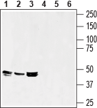
 Western blot analysis of rat spleen (lanes 1 and 4), mouse spleen (lanes 2 and 5) and mouse brain (lanes 3 and 6) lysates:1-3. Anti-Septin-7 Antibody (#AIP-047), (1:200).
4-6. Anti-Septin-7 Antibody, preincubated with Septin-7 Blocking Peptide (#BLP-IP047) (#BLP-IP047).
Septin 7 is a highly conserved GTP-binding filamentous protein. Septins are important for the organization of the actin cytoskeleton. Septin7 also plays an important role in cytokinesis, and membrane dynamics1,2.
Septins were first discovered in yeast and since, 13 septin genes have been identified in mammals (SEPT1 to SEPT12 and SEPT14). The 13 gene-encoded proteins can be classified into four distinct subgroups based on their sequence homology of their domain structure2.
Septins share a highly conserved structure composed of the central GTPase domain and N- and C-terminal domains of various lengths3. They self-assemble into higher-order structures, including filaments and rings in tri-, hexa-, or octameric complexes consisting of multiple septin polypeptides, which are typical for different cell types. The various septin isoforms seem to determine the function and regulation of the septin complex4.
Septin-7 regulates Ca2+ entry through Orai1 by regulating the opening of the channel5.
Dysregulation of septins leads to human diseases such as neurodegenerative and bleeding disorders. In non-dividing cells such as neuronal tissue and platelets septins have been associated with exocytosis1,2.
