Overview
- Peptide (C)RSQEGRQESRSDKAK, corresponding to amino acid residues 614-628 of rat Shank1 (Accession Q9WV48). Intracellular, between the SH3 and PDZ domains.

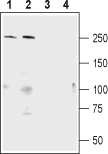
 Western blot analysis of mouse (lanes 1 and 3) and rat (lanes 2 and 4) brain lysate:1,2. Anti-Shank1 Antibody (#APZ-011), (1:400).
3,4. Anti-Shank1 Antibody, preincubated with Shank1 Blocking Peptide (#BLP-PZ011).
 Expression of Shank1 in rat hippocampusImmunohistochemical staining of perfusion-fixed frozen rat brain sections using Anti-Shank1 Antibody (#APZ-011), (1:300), followed by goat anti-rabbit-AlexaFluor-488 secondary antibody. Shank1 staining (green) is detected in interneurons (vertical arrows) and in the pyramidal layer (P, horizontal arrows). Nuclei are stained with DAPI (blue).
- Jiang, Y. H. and Ehlers, M.D. (2013) Neuron 78, 8.
- Sheng, M. and Kim, E. (2000) J. Cell Sci. 113, 1851.
- Stella, S.L. Jr. et al. (2012) PLoS One 7, e43463.
- Guilmatre, A. et al. (2014) Dev. Neurobiol. 74, 113.
- Lim, S. et al. (1999) J. Biol. Chem. 274, 29510.
- Lennertz, L. et al. (2012) Eur. Arch. Psychiatry Clin. Neurosci. 262, 117.
- Hung, A.Y. et al. (2008) J. Neurosci. 28, 1697.
Shank family proteins (shank1, shank2 and shank3) are neuronal scaffold proteins that organize a cytoskeleton-associated signaling complex at the postsynaptic density (PSD) of nearly all excitatory glutamatergic synapses in the mammalian brain1.
The Shank family, including Shank1, has five conserved protein domains: 1. ankyrin repeat domain (ANK), 2. Src homology 3 (SH3) domain, 3. PSD-95/Discs large/ZO-1 (PDZ) domain, 4. proline-rich region containing homer and cortactin-binding sites (Pro), 5. sterile α motif (SAM) domain2. All these domains can act as sites for specific protein-protein interactions, and the presence of these multiple domains within a single polypeptide is consistent with a function as a scaffolding protein. Shanks interact directly or indirectly with all major types of glutamate receptors: NMDA receptors, AMPA receptors, and mGluRs via different domains. Shank proteins also interact with several actin regulatory proteins and L-type Ca2+ (CaV1.3) channels in medium spiny neurons of the striatum3.
Shank1 mRNA, being synthesized in dendritic spine, can locally control the spine morphology. The Shank1 gene is located on chromosome 19q13.33 and contains 23 exons and two alternative promoters leading to two distinct isoforms4. Shank1 is highly expressed in the cortex and hippocampus5.
Recent studies indicate that Shank1 is a causative gene for idiopathic autism spectrum disorders (ASD)1. In addition it was found that Shank1 has a role in working memory deficits in schizophrenia6. Shank1 knock-out mice display increased anxiety and decreased vocal communication7.
Application key:
Species reactivity key:
Alomone Labs is pleased to offer a highly specific antibody directed against an epitope of rat Shank1. Anti-Shank1 Antibody (#APZ-011) can be used in western blot and immunohistochemistry applications. It has been designed to recognize Shank1 from human, rat, and mouse samples.
