Overview
- Peptide (C)SDKRGLGLQVRDQFR, corresponding to amino acid residues 412 - 426 of mouse SLC19A1 (Accession P41438). 6th extracellular loop.

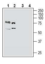
 Western blot analysis of rat brain membranes (lanes 1 and 5), mouse brain membranes (lanes 2 and 6), rat small intestine lysate (lanes 3 and 7) and rat liver membranes (lanes 4 and 8):1-4. Anti-SLC19A1/RFC1 (extracellular) Antibody (#AFT-001), (1:400).
5-8. Anti-SLC19A1/RFC1 (extracellular) Antibody, preincubated with SLC19A1/RFC1 (extracellular) Blocking Peptide (#BLP-FT001). Western blot analysis of human MCF-7 breast adenocarcinoma cell line lysate (lanes 1 and 3) and human K562 myelogenous leukemia cell line lysate (lanes 2 and 4):1, 2. Anti-SLC19A1/RFC1 (extracellular) Antibody (#AFT-001), (1:400).
3, 4. Anti-SLC19A1/RFC1 (extracellular) Antibody, preincubated with SLC19A1/RFC1 (extracellular) Blocking Peptide (#BLP-FT001).
 Expression of SLC19A1 in rat hippocampus.Immunohistochemical staining of perfusion-fixed frozen rat brain sections with Anti-SLC19A1/RFC1 (extracellular) Antibody (#AFT-001), (1:1200), followed by goat anti-rabbit-AlexaFluor-488. A. Staining in the rat hippocampal CA3 region, showed immunoreactivity (green) in neuronal profiles in the pyramidal layer (arrows). B. Pre-incubation of the antibody with SLC19A1/RFC1 (extracellular) Blocking Peptide (BLP-FT001), suppressed staining. Cell nuclei are stained with DAPI (blue). P = pyramidal layer.
- Prasad, P.D. et al. (1995) Biochem. Biophys. Res. Commun. 206, 681.
- Yee, S.W. et al. (2011) Pharmacogenet. Genomics 20, 708.
- Cao, W. and Matherly, L.H. (2004) Biochem. J. 378, 201.
- Ifergan, I. et al. (2008) J. Biol. Chem. 283, 20687.
- Whetstine, J.R. et al. (2002) Biochem. J. 367, 629.
- Hu, Y.H. et al. (2019) Curr. Pharm. Des. 25, 627.
Solute Carrier Transporter 19A1, (SLC19A1, RFC1) is a high-capacity, bi-directional transporter of 5-methyl-tetrahydrofolate and thiamine monophosphate1,2. SLC19A1 is characterized by 12 transmembrane domains (TMDs) and cytoplasm oriented N- and C-termini3.
Folate is a major nutrient that supports important physiological functions such as DNA synthesis, cell division and substrate methylation. Low folate levels caused by suboptimal intake, transport and cellular utilization of folate, is a risk factor for diseases such as spina bifida, cardiovascular diseases and certain types of cancer1.
SLC19A1 is the main folate transporter and it plays a critical role in folate uptake and homeostasis in mammalian cells4. High SLC19A1 transcript levels are detected in liver and placenta, with appreciable levels in other tissues, including kidney, lung, bone marrow, intestine, brain, and portions of the central nervous system5.
SLC19A1 is also involved in the uptake of Methotrexate (MTX); one of the leading chemotherapeutic agents with the best demonstrated efficacies against childhood acute lymphoblastic leukemia and hence malfunction of this transporter also effects the efficiency of this type of cancer treatment6.
Application key:
Species reactivity key:
Anti-SLC19A1/RFC1 (extracellular) Antibody (#AFT-001) is a highly specific antibody directed against an epitope of the mouse protein. The antibody can be used in western blot and live cell flow cytometry applications. It has been designed to recognize SLC19A1 from human, rat, and mouse samples.

