Overview
- Peptide (C)GSNQTEPYYDMTSN, corresponding to amino acid residues 30-43 of rat SSTR2 (Accession P30680). Extracellular, N-terminus.

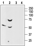
 Western blot analysis of rat brain (lanes 1 and 3) and pancreas (lanes 2 and 4) lysates:1-2. Anti-Somatostatin Receptor Type 2 (extracellular) Antibody (#ASR-006), (1:200).
3-4. Anti-Somatostatin Receptor Type 2 (extracellular) Antibody, preincubated with Somatostatin Receptor Type 2 (extracellular) Blocking Peptide (#BLP-SR006).
 Expression of SSTR2 in rat hippocampusImmunohistochemical staining of rat hippocampus using Anti-Somatostatin Receptor Type 2 (extracellular) Antibody (#ASR-006). A. SSTR2 appears in neural processes (green) that are perpendicular to dentate granule layer (arrow). B. Staining of axons with mouse anti-Neurofilament 200 (NF200, red). C. Confocal merge of SSTR2 and NF200 images suggests that SSTR2 is not present in axons that run through the granule layer and hilus, but rather in neuronal dendrites that ascend toward the dentate hilus (arrow).
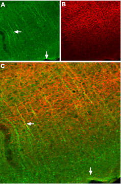
 Expression of SSTR2 in mouse cortexImmunohistochemical staining of frozen mouse cortex sections using Anti-Somatostatin Receptor Type 2 (extracellular) Antibody (#ASR-006), (1:100). A. SSTR2 appears in neural processes (green, horizontal arrow) that are perpendicular to the cortical surface (vertical arrow). B. Staining of axons with mouse anti-Neurofilament 200 (NF200, red) appears mostly in the deep layers. C. Confocal merge of SSTR2 and NF200 images suggests that SSTR2 is not present in axons that run in the deep layers, but rather in neuronal dendrites that ascend toward upper layers of cortex (vertical arrow).
 Expression of SSTR2 in human HT-29 cellsCell surface detection of SSTR2 in intact living human colorectal adenocarcinoma (HT-29) cells. A. Extracellular staining of cells with Anti-Somatostatin Receptor Type 2 (extracellular) Antibody (#ASR-006), (1:50), (red). B. Merged image of A with live view of cell.
- Hofland, L.J. and Lamberts, S.W. (2001) Ann. Oncol. 12, S31.
- Fombonne, J. et al. (2003) Rep. Biol. Endocrinol. 1, 19.
- Slooter, G.D. et al. (2001) Br. J. Surg. 88, 31.
- Schulz, S. et al. (2002) Gyn. Oncol. 84, 235.
Somatostatin is a small cyclic peptide that is widely expressed throughout the central nervous system and peripheral tissues.1 In peripheral tissues, somatostatin exerts inhibitory effects on secretion processes, whereas in the brain, it acts as a neurotransmitter in both a stimulatory and an inhibitory manner.1,2
Somatostatin mediates its action via six high affinity G-protein coupled receptors (SSTR1, SSTR2a, SSTR2b, SSTR3, SSTR4, and SSTR5), which are encoded by five genes.1,2 Expression of the different receptors is developmentally regulated in a time- and tissue-specific manner.2
Somatostatin receptors have been found on a variety of neuroendocrine tumors, such as paragangliomas, carcinoids, and breast tumors.3 Synthetic peptide derivatives of somatostatin have been successfully used in the treatment of neuroendocrine malignancies and in vivo imaging of tumors that are positive for somatostatin receptors.4
In general, SSTR2 is the most common SSTR subtype found in human tumors, followed by SSTR1, with SSTR3 and SSTR4 being less common.
Application key:
Species reactivity key:
Alomone Labs is pleased to offer a highly specific antibody directed against the N-terminal domain of the rat SSTR2. Anti-Somatostatin Receptor Type 2 (extracellular) Antibody (#ASR-006), can be used in western blot, immunohistochemistry and immunocytochemistry applications. It has been designed to recognize SSTR2 from mouse, human and rat samples.

