Overview
- Peptide (C)KRRTPHAT(S)FNGED, corresponding to amino acid residues 652 - 665 of human SORL1 (Accession Q92673). Extracellular, N-terminus.

 Western blot analysis of rat brain membranes (lanes 1 and 3) and mouse brain membranes (lanes 2 and 4):1, 2. Anti-SORL1 (extracellular) Antibody (#ANR-132), (1:200).
3, 4. Anti-SORL1 (extracellular) Antibody, preincubated with SORL1 (extracellular) Blocking Peptide (#BLP-NR132). Western blot analysis of human Jurkat T-cell leukemia cell line lysate (lanes 1 and 5), human SH-SY5Y neuroblastoma cell line lysate (lanes 2 and 6), human Colo-205 colon adenocarcinoma cell line lysate (lanes 3 and 7) and HepG2 hepatocellular carcinoma (lanes 4 and 8):1-4. Anti-SORL1 (extracellular) Antibody (#ANR-132), (1:200).
5-8. Anti-SORL1 (extracellular) Antibody, preincubated with SORL1 (extracellular) Blocking Peptide (#BLP-NR132).
 Expression of SORL1 in rat parietal cortex.Immunohistochemical staining of perfusion-fixed frozen rat brain sections with Anti-SORL1 (extracellular) Antibody (#ANR-132), (1:300), followed by goat anti-rabbit-AlexaFluor-488. A. SORL1 immunoreactivity (green) appears in neuronal profiles in layer III (arrows). B. Pre-incubation of the antibody with SORL1 (extracellular) Blocking Peptide (BLP-NR132), suppressed staining. Cell nuclei are stained with DAPI (blue).
 Expression of SORL1 in mouse hippocampus.Immunohistochemical staining of perfusion-fixed frozen mouse brain sections with Anti-SORL1 (extracellular) Antibody (#ANR-132), (1:300), followed by goat anti-rabbit-AlexaFluor-488. A. Staining in mouse hippocampal CA1 region revealed SORL1 immunoreactivity (green) in neuronal profiles in the pyramidal layer (P) and in the stratum oriens (SO, arrows). B. Pre-incubation of the antibody with SORL1 (extracellular) Blocking Peptide (BLP-NR132), suppressed staining. Cell nuclei are stained with DAPI (blue). P = pyramidal layer, SO = stratum oriens, SR = stratum radiatum.
- Barthelson, K. et al. (2020) J. Alzheimers Dis. Rep. 4, 123.
- Motoi, Y. et al. (1999) Brain Res. 833, 209.
- Jacobsen, L. et al. (1996) J. Biol. Chem. 271, 31379.
Sortilin-related receptor1 (SORL1, also known as LR11 or SORLA) is a receptor involved in intracellular sorting and trafficking of proteins into their respective subcellular compartments1.
Direction of proteins into their proper compartments is essential for normal cell function and defects in these pathways are thought to be important factors in the pathogenesis of Alzheimer’s disease (AD).
The full-length transcript of SORL1 encodes a 250 kDa, membrane-bound protein comprised of multiple functional domains. These include a vacuolar protein sorting 10 (VPS10) domain, five low-density lipoprotein receptor (LDLR) class B repeats, an epidermal growth factor-like (EGF-like) domain, eleven LDLR class A repeats, six fibronectin-type (FN-type) repeats, a transmembrane domain and a cytosolic domain containing recognition motifs for cytosolic adaptors1.
SORL1 is widely expressed in the human brain, particularly in neurons of the hippocampus and some nuclei of the brainstem and Purkinje cells, and has slightly weaker expression in neurons of the thalamus and the hypothalamus2. It is also expressed in other tissue types such as the testes, ovaries, thyroid, and lymph nodes3.
Application key:
Species reactivity key:
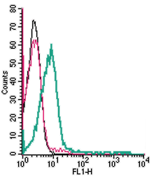
Anti-SORL1 (extracellular) Antibody (#ANR-132) is a highly specific antibody directed against an epitope of the human protein. The antibody can be used in western blot and live cell flow cytometry applications. It has been designed to recognize SORL1 from human, rat, and mouse samples.

