Overview
- Peptide (C)KHWGRRKAW(S)RQLGE, corresponding to amino acid residues 42-56 of mouse TREM2 (Accession Q99NH8). Extracellular, N-terminus.

 Western blot analysis of mouse (lanes 1 and 3) and rat (lanes 2 and 4) brain lysates:1,2. Anti-TREM2 (extracellular) Antibody (#ANR-018), (1:200).
3,4. Anti-TREM2 (extracellular) Antibody, preincubated with TREM2 (extracellular) Blocking Peptide (#BLP-NR018). Western blot analysis of mouse BV-2 microglia (lanes 1 and 4), human U-87 MG glioblastoma (lanes 2 and 5) and human THP-1 monocytic leukemia (lanes 3 and 6) cell line lysates:1-3. Anti-TREM2 (extracellular) Antibody (#ANR-018), (1:200).
4-6. Anti-TREM2 (extracellular) Antibody, preincubated with TREM2 (extracellular) Blocking Peptide (#BLP-NR018).
 Expression of TREM2 in mouse hippocampus in a kainic acid neurodegeneration model.Immunohistochemical staining of perfusion-fixed frozen mouse brain sections collected 4 days post kainate injection with Anti-TREM2 (extracellular) Antibody (#ANR-018), (1:300), followed by goat anti-rabbit-AlexaFluor-488. A. Staining in the mouse hippocampal CA1 region showed TREM2 immunoreactivity (green) in astrocytes in the stratum oriens (SO) and the stratum radiatum (SR). B. Pre-incubation of the antibody with TREM2 (extracellular) Blocking Peptide (BLP-NR018), suppressed staining. Cell nuclei are stained with DAPI (blue).
- Shirotani, K. et al. (2019) Sci. Rep. 9, 7508.
- Del-Aguila, J.L. et al. (2019) Mol. Neurodegener. 14, 18.
- Zhong, L. et al. (2019) Nat. Commun. 10, 1365.
TREM2 (triggering receptor expressed on myeloid cells-2) is a type I transmembrane microglial surface receptor that is found to be expressed on myeloid cells including monocyte-derived dendritic cells, osteoclasts and bone-marrow derived macrophage. TREM2 protein transduces intracellular signals that regulate microglial functions such as cytokine production, migration, proliferation, phagocytosis, cell survival, synapse elimination, and compaction of amyloid plaques1,2.
The TREM2 structure includes an immunoglobulin-like extracellular domain responsible for ligand binding, a transmembrane region and a short cytoplasmatic tail. The protein also includes an ectodomain that binds anionic and zwitterionic lipids, apolipoproteins and lipoprotein particles2,3.
Studies show that TREM2 knockout mice display decreased levels of inflammatory cytokines. TREM2 has been shown to control two signaling pathways in the brain: regulation of phagocytosis and suppression of inflammatory reactivity. Nasu-Hakola disease, a rare and fatal disease, and polycystic lipomembranous osteodysplasia with sclerosing leukoencephalopathy has been found to be linked to homozygous loss-of-function mutations in TREM2 protein1,2. Studies also show that variants of TREM2 are associated with Alzheimer’s disease2,3.
Application key:
Species reactivity key:
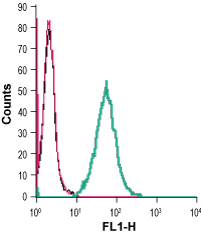
Anti-TREM2 (extracellular) Antibody (#ANR-018) is a highly selective antibody directed against an epitope of the mouse protein. The antibody can be used in western blot, immunohistochemistry and live cell flow cytometry applications. The antibody recognizes an extracellular epitope and can be used to detect the protein in living cells. It has been designed to recognize TREM2 from mouse, human, and rat samples.

