Overview
- Peptide (C)NSTGIINETSDHSE, corresponding to amino acid residues 747-760 of human TRPA1 (Accession O75762). 1st extracellular loop.
- Rat DRG lysate (1:200). Human urethra (1:2000) (Gratzke, C. et al. (2009) Eur. Urol. 55, 696.).
 Western blot analysis of rat DRG (lanes 1,2), non-differentiated PC12 cells (lanes 3,5) and differentiated PC12 cells (lanes 4,6) lysates:1,3,4. Anti-TRPA1 (extracellular) Antibody (#ACC-037), (1:200).
2,5,6. Anti-TRPA1 (extracellular) Antibody, preincubated with TRPA1 (extracellular) Blocking Peptide (#BLP-CC037).
- Differentiated PC12 cell lines (6.5 μg).
- Mouse and rat DRG frozen sections (1:200). Human urethra (1:500) (Gratzke, C. et al. (2009) Eur. Urol. 55, 696.).
- Rat intact living PC12 cells (1:50).
The TRPA family is comprised of only one mammalian member, the TRPA1 (formerly named ANKTM1). TRPA1 is expressed in peripheral sensory neurons, where it is suggested to contribute to the detection of painful stimuli.1
Originally, it was thought that TRPA channels sensed painfully cold temperatures,2 but a more conservative description is that TRPA1 is sensitive to membrane/cytoskeletal perturbations caused by low temperatures3-5 and perhaps stretch.6 In addition, it is sensitive to pungent natural compounds present in cinnamon oil, mustard oil, and wintergreen oil.
TRPA1 is also expressed in hair cells, where its role in sensing mechanical forces is still unclear and controversial.1
TRPA1 has a similar structure to all other TRP ion channels; six transmembrane domains, intracellular N-and C-terminus. However, the N-terminal domain possesses 17 ankyrin repeats that might indicate its potential role as a mechanosensor.6,7
In addition, TRPA1 is expressed in nociceptive neurons expressing TRPV1 and might serve as a marker for polymodal nociceptors.8
Application key:
Species reactivity key:

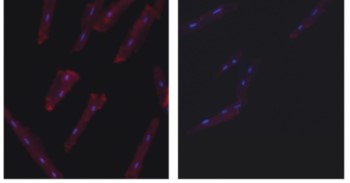
Knockout validation of Anti-TRPA1 (extracellular) Antibody in mouse cardiomyocytes.Immunocytochemical staining of mouse cardiomyocytes using Anti-TRPA1 (extracellular) Antibody (#ACC-037). TRPA1 staining (red) is detected in cardiomyocytes from wild type mice (left panel). Lack of TRPA1 immunoreactivity is observed in TRPA1-/- cells (right panel).Adapted from Conklin, D.J. et al. (2019) Am. J. Physiol. 316, H889. with permission of The American Physiological Society.
