Overview
Application key:
Species reactivity key:

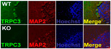
Knockout validation of Anti-TRPC3 Antibody in mouse cerebellumImmunohistochemical staining of mouse cerebellum sections using Anti-TRPC3 Antibody (#ACC-016). TRPC3 staining (green) is detected in neurons and partially overlaps with the neuronal marker MAP-2 (red). No staining for TRPC3 is observed in TRPC3-/- mice (lower panels).Adapted from Feng, S. et al. (2013) Proc. Natl. Acad. Sci. U.S.A. 110, 11011. with permission of the National Academy of Sciences of the United States of America.
Specifications
- Peptide HKLSEKLNPSVLRC, corresponding to amino acid residues 822-835 of mouse TRPC3 (Accession Q9QZC1). Intracellular, C-terminus.
Applications
- Rat heart membranes (1:200-1:1000). Addition of 0.1-0.5% Tween-20 to the antibody solution may strengthen the signal.
 Western blot analysis of rat heart membranes:1. Anti-TRPC3 Antibody (#ACC-016), (1:200).
2. Anti-TRPC3 Antibody, preincubated with TRPC3 Blocking Peptide (#BLP-CC016).
- HEK-293 cells transfected with human TRPC3 (Kwan, H.Y. et al. (2004) Proc. Natl. Acad. Sci. U.S.A. 101, 2625.).
- Mouse and rat brain frozen sections. Rat pituitary gland paraffin embedded section (1:100). Rat cerebellum frozen section.
- C6 cell line (rat brain glioma), (1:500).
- Rat microglia cells (Mizoguchi, Y. et al. (2014) J. Biol. Chem. 289, 18549.).
Scientific Background
The Transient Receptor Potential (TRP) superfamily is one of the largest ion channel families and consists of diverse groups of proteins. In mammals, about 28 genes encode the TRP ion channel subunits. The mammalian TRP superfamily comprises six subfamilies known as the TRPC (canonical), TRPV (vanilloid), TRPM (melastatin), TRPML (mucolipins), TRPP (polycystin) and the TRPA (ANKTM1) ion channels.1-4
The TRPC subfamily consists of seven proteins named TRPC1 to 7 which can be further divided into four subgroups based on their sequence homology and functional similarities:
1. TRPC1
2. TRPC4 and TRPC5
3. TRPC3, TRPC6, TRPC7
4. TRPC2.2,5
They are highly expressed in the central nervous system and to a lesser extent in peripheral tissues.
TRPC3, TRPC6 and TRPC7 form non-selective cationic channels that are activated by the stimulation of GPCRs.
Different modes of activation, DAG and SOC, were described for the TRPC3 channel by different investigators working with heterologous expression systems.6,7
