Overview
- Peptide HKLSEKLNPSVLRC, corresponding to amino acid residues 822-835 of mouse TRPC3 (Accession Q9QZC1). Intracellular, C-terminus.

 Expression of TRPC3 in rat DRGImmunohistochemical staining of rat dorsal root ganglion (DRG) frozen sections using Anti-TRPC3-ATTO Fluor-594 Antibody (#ACC-016-AR), (1:50). Staining is present in neuronal cell bodies. Hoechst 33342 is used as the counterstain.
 Colocalization of TRPC6 and TRPC3 in rat cerebellumImmunohistochemical staining of rat cerebellum frozen section using Guinea pig Anti-TRPC6 Antibody (#ACC-017-GP) and rabbit Anti-TRPC3-ATTO Fluor-594 Antibody (#ACC-016-AR). A. TRPC6 staining (green) appears in molecular layer and in Purkinje cells. B. In the same section as in A, staining of TRPC3 (red) appears as well in both molecular layer and Purkinje cells. C. Merge images of A and B indicates co-localization in Purkinje cells and molecular layer.
 Multiplex staining of VMAT2 and TRPC3 in rat brainImmunohistochemical staining of perfusion-fixed frozen rat substantia nigra sections using Anti-VMAT2-ATTO Fluor-488 Antibody (#AMT-006-AG), (1:60) and Anti-TRPC3-ATTO Fluor-594 Antibody (#ACC-016-AR), (1:60). A. VMAT2 staining (green). B. The same section stained for TRPC3 (red). C. Merged image demonstrates the ubiquitous colocalization of VMAT2 and TRPC3 in cells of the substantia nigra pars compacta. Arrows point at examples of co-expression.
- Moran, M.M. et al. (2004) Current Opin. Neurobiol. 14, 362.
- Clapham, D.E. et al. (2003) Pharmacol. Rev. 55, 591.
- Clapham, D.E. (2003) Nature 426, 517.
- Padinjat, R. and Andrews, S. (2004) J. Cell. Sci. 117, 5707.
- Huang, C.L. (2004) J. Am. Soc. Nephrol. 15, 1690.
- Vazquez, G. et al. (2003) J. Biol.Chem. 278, 21649.
- Venkatachalam, K. et al. (2003) J. Biol.Chem. 278, 29031.
The Transient Receptor Potential (TRP) superfamily is one of the largest ion channel family and consists of diverse groups of proteins. In mammals about 28 genes encode the TRP ion channel subunits. The mammalian TRP superfamily comprises six subfamilies known as the TRPC (canonical), TRPV (vanilloid), TRPM (melastatin), TRPML (mucolipins), TRPP (polycystin) and the TRPA (ANKTM1) ion channels.1-4
The TRPC subfamily consists of seven proteins named TRPC1 to 7 which can be further divided into four subgroups based on their sequence homology and functional similarities: TRPC1; TRPC4 and TRPC5; TRPC3, TRPC6, TRPC7 and TRPC2.2,5 They are highly expressed in the central nervous system and to a lesser extent in peripheral tissues.
TRPC3, TRPC6 and TRPC7 form non-selective cationic channels that are activated by the stimulation of GPCRs.
TRPC3 was shown to be activated by DAG and SOC, in heterologous expression systems.6,7
Application key:
Species reactivity key:
Anti-TRPC3 Antibody (#ACC-016) is a highly specific antibody directed against an epitope of the mouse protein. The antibody can be used in western blot, immunoprecipitation, immunohistochemistry, immunocytochemistry, and indirect flow cytometry applications. It has been designed to recognize TRPC3 from mouse, rat, and human samples.
Anti-TRPC3-ATTO Fluor-594 Antibody (#ACC-016-AR) is directly labeled with ATTO-594 fluorescent dye. ATTO dyes are characterized by strong absorption (high extinction coefficient), high fluorescence quantum yield, and high photo-stability. The ATTO-594 fluorescent label belongs to the class of Rhodamine dyes and can be used with fluorescent equipment typically optimized to detect Texas Red and Alexa-594. Anti-TRPC3-ATTO Fluor-594 Antibody can be used in immunohistochemistry applications and is specially suited to experiments requiring simultaneous labeling of different markers.

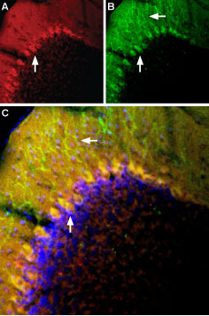
Multiplex staining of mGluR1 and TRPC3 in mouse cerebellumImmunohistochemical staining of perfusion-fixed frozen mouse cerebellum sections using Anti-TRPC3-ATTO Fluor-594 Antibody (#ACC-016-AR), (1:60) and Anti-mGluR1 (extracellular)-ATTO Fluor-488 Antibody (#AGC-006-AG), (1:60). A. TRPC3 staining (red). B. mGluR1 staining (green). C. Merge of the two images suggests extensive co-localization in Purkinje cells (vertical arrows). Note expression of mGluR1 in Purkinje dendrites (horizontal arrow) but not of TRPC3. Cell nuclei are stained with DAPI (blue).
Applications
Citations
- Western blot analysis of mouse kidney lysate using #ACC-016. Tested in TRPC3/6/7-/- mice.
Liu, B. et al. (2017) Am. J. Transl. Res. 9, 5619. - Western blot analysis of mouse brain lysate and immunohistochemical staining of mouse cerebellum using #ACC-016. Tested in Trpc3-/- mice.
Feng, S. et al. (2013) Proc. Natl. Acad. Sci. U.S.A. 110, 11011.
