Overview
- Peptide (C)KEKHAHEEDSSIDYDL, corresponding to amino acid residues 943-958 of mouse TRPC4 (Accession Q9QUQ5). Intracellular, C-terminus.

 Western blot analysis of rat brain membranes:1. Anti-TRPC4 Antibody (#ACC-018), (1:200).
2. Anti-TRPC4 Antibody, preincubated with TRPC4 Blocking Peptide (#BLP-CC018). Western blot analysis of human PC3 Caucasian prostate adenocarcinoma (lanes 1 and 3) and human LNCaP prostate carcinoma (lanes 2 and 4) cell lysates:1,3. Anti-TRPC4 Antibody (#ACC-018), (1:200).
2,4. Anti-TRPC4 Antibody, preincubated with TRPC4 Blocking Peptide (#BLP-CC018).
 Immunoprecipitation of rat brain lysate:1. Rat brain lysate.
2. Lysate immunoprecipitated with Anti-TRPC4 Antibody (#ACC-018), (6 µg).
3. Lysate immunoprecipitated with pre-immune rabbit serum.
The upper arrow indicates the TRPC4 channel while the lower arrow indicates the IgG heavy chain.
Western blot analysis was performed with Anti-TRPC4 Antibody.
 Expression of TRPC4 in mouse cerebellumImmunohistochemical staining of mouse cerebellum frozen sections with Anti-TRPC4 Antibody (#ACC-018). A. TRPC4 (red) appears in Purkinje cells (arrows) and in the molecular (Mol) layer. B. Staining with mouse anti-parvalbumin (PV) in the same brain section. C. Confocal merge of TRPC4 and PV demonstrates partial co-localization in the Purkinje and the molecular layers.
- Rat type I astrocytes (1:25) (Grimaldi, M. et al. (2003) J. Neurosci. 23, 4737.).
Human U373 MG cells (1:100) (Barajas, M. et al. (2008) J. Neurosci. Res. 86, 3456.).
- Moran, M.M. et al. (2004) Current Opin. Neurobiol. 14, 362.
- Clapham, D.E. et al. (2003) Pharmacol. Rev. 55, 591.
- Clapham, D.E. (2003) Nature 426, 517.
- Padinjat, R. and Andrews, S. (2004) J. Cell. Sci. 117, 5707.
- Huang, C.L. (2004) J. Am. Soc. Nephrol. 15, 1690.
- Liu, X. et al. (2003) J. Biol.Chem. 278, 11337.
The Transient Receptor Potential (TRP) superfamily is one of the largest ion channel families and consists of diverse groups of proteins. In mammals, about 28 genes encode the TRP ion channel subunits. The mammalian TRP superfamily comprises six subfamilies known as the TRPC (canonical), TRPV (vanilloid), TRPM (melastatin), TRPML (mucolipins), TRPP (polycystin) and the TRPA (ANKTM1) ion channels.1-4
The TRPC subfamily consists of seven proteins named TRPC1 to 7 which can be further divided into four subgroups based on their sequence homology and functional similarities:
1. TRPC1
2. TRPC4 and TRPC5
3. TRPC3, TRPC6, TRPC7
4. TRPC2.2,5
They are highly expressed in the central nervous system and to a lesser extent in peripheral tissues.
TRPC4 can form heterotetramers with TRPC1. TRPC4, TRPC1 and TRPC5 can be activated either by calcium store depletion or by GPCR stimulation pathways and are also assumed to form components of store operated channels in some cell types such as salivary gland cells, endothelial cells and vascular smooth muscle cells. 6
Application key:
Species reactivity key:
Anti-TRPC4 Antibody (#ACC-018) is a highly specific antibody directed against an epitope of the mouse protein. The antibody can be used in western blot, immunoprecipitation, immunohistochemistry, and immunocytochemistry applications. It has been designed to recognize TRPC4 from human, mouse, and rat samples.

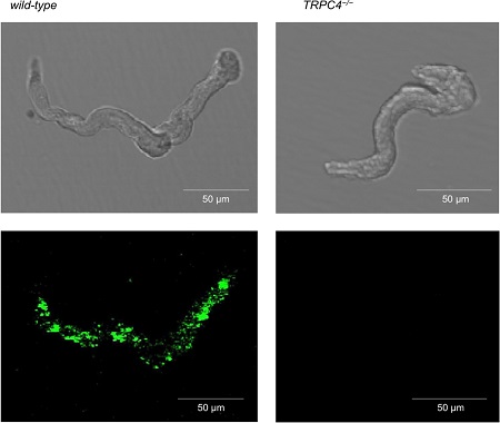
Knockout validation of Anti-TRPC4 Antibody in mouse smooth muscle cells.Immunocytochemical staining of mouse smooth muscle cells with Anti-TRPC4 Antibody (#ACC-018). TRPC4 staining is prominent in wild type cells (left panels). TRPC4 is not detected in TRPC4-/- cells (right panels) demonstrating the specificity of the antibody.Adapted from Griffin, C.S. et al. (2018) Sci. Rep. 8, 9264. with permission of SPRINGER NATURE.
Applications
Citations
 Expression of TRPC4 in human MEC-1 cells.Immunocytochemical staining of human microvascular endothelial cells (HMEC-1) using Anti-TRPC4 Antibody (#ACC-018).
Adapted from Graziani, A. et al. (2010) J. Biol. Chem. 285, 4213. with permission of The American Society for Biochemistry and Molecular Biology.
- Immunocytochemical staining of mouse smooth muscle cells. Also tested in TRPC4-/- cells.
Griffin, C.S. et al. (2018) Sci. Rep. 8, 9264. - Western blot analysis of rat pulmonary artery lysates. Also tested in siRNA treated cells.
Jiang, H.N. et al. (2016) Biomed. Pharmacol. 82, 20.
- Rat pulmonary artery lysates. Also tested in siRNA treated cells.
Jiang, H.N. et al. (2016) Biomed. Pharmacol. 82, 20. - Rat neonatal ventricular cardiomyocytes (1:200).
Sabourin, J. et al. (2016) J. Biol. Chem. 291, 13394. - Rat INS-1 cell lysate.
Park, S.H. et al. (2013) Proc. Natl. Acad. Sci. U.S.A. 110, 12673. - Mouse brain lysate.
Feng, S. et al. (2013) Proc. Natl. Acad. Sci. U.S.A. 110, 11011. - Human primary myotubes.
Harisseh, R. et al. (2013) Am. J. Physiol. 304, C881. - Rat distal pulmonary smooth muscle cell lysate (PASMCs).
Zhang, Y. et al. (2013) Am. J. Physiol. 304, C833.
- Rat neonatal ventricular cardiomyocytes.
Sabourin, J. et al. (2016) J. Biol. Chem. 291, 13394.
- Mouse smooth muscle cells. Also tested in TRPC4-/- cells.
Griffin, C.S. et al. (2018) Sci. Rep. 8, 9264. - Rat primary cultured fetal and neonatal ventricular myocytes (1:100).
Jiang, Y. et al. (2014) Cell Tissue Res. 355, 201. - Human primary myotubes.
Harisseh, R. et al. (2013) Am. J. Physiol. 304, C881. - Human U373 MG cells (1:100).
Barajas, M. et al. (2008) J. Neurosci. Res. 86, 3456. - Rat type I astrocytes (1:25).
Grimaldi, M. et al. (2003) J. Neurosci. 23, 4737.
- Kadeba, P.I. et al. (2013) Cell Calcium 53, 275.
- Graziani, A. et al. (2010) J. Biol. Chem. 285, 4213.
- Facemire, C.S. et al . (2004) Am. J. Physiol. 286, F546.
- Castellano, L.E. et al. (2003) FEBS Lett. 541, 69.
- Vandebrouck, C. et al. (2002) J. Cell. Biol. 158, 1089.
- Wu, X. et al. (2002) J. Biol. Chem. 277, 13597.
