Overview
- Peptide EKEQSWIPKIFKK(C), corresponding to amino acid residues 5-17 of human TRPM4 (Accession Q8TD43). Intracellular, N-terminus.

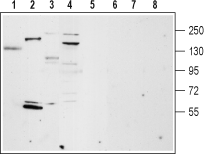 Western blot analysis of rat lung (lanes 1 and 5), rat heart membrane (lanes 2 and 6), mouse brain membrane (lanes 3 and 7) and ms1 mouse pancreas cells lysate (lanes 4 and 8):1-4. Anti-TRPM4 Antibody (#ACC-044), (1:200).
5-8. Anti-TRPM4 Antibody, preincubated with TRPM4 Blocking Peptide (#BLP-CC044).
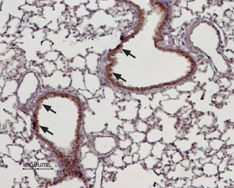 Expression of TRPM4 in rat lungImmunohistochemical staining of rat lung paraffin embedded sections using Anti-TRPM4 Antibody (#ACC-044), (1:100). TRPM4 is expressed in the respiratory epithelium of the bronchioli (arrows). Hematoxilin is used as the counterstain.
- Fleig, A. et al. (2004) Trends. Pharmacol. Sci. 25, 633.
- Kraft, R. et al. (2005) Pflugers. Arch. 451, 204.
- Huang, C.L. (2004) J. Am. Soc. Nephrol. 15, 1690.
- Vazquez, E. et al. (2006) Semin. Cell Dev. Biol. 17, 607.
The mammalian melastatin-related transient receptor potential (TRPM) is a subfamily of the TRP family. The family was named after the first discovered member, melastatin (TRPM1) whose gene was identified in metastatic and benign melanomas1-3.
The TRPM family consists of eight members designated as TRPM1-8 that can be further divided into four pairs: TRPM1 and TRPM3; TRPM2 and TRPM8; TRPM4 and TRPM5; and TRPM6 and TRPM71,2.
The TRPM proteins share structural homology with other members of the TRP superfamily channels; six putative transmembrane domains, and cytoplasmic N- and C-termini. However, due to their long N- and C-termini they are also named the long TRP channel family1-3.
TRPM4 and TRPM5 are the only TRP channels that are permeable to monovalent cations but not to Ca2+.
TRPM4 is expressed as at least two alternative spliced isoforms, TRPM4a and TRPM4b which consist of shorter and longer N-terminal regions, respectively. TRPM4a displays little activity, TRPM4b and TRPM5 appear to be directly activated by an increase in intracellular Ca2+ concentration4.
TRPM4 is expressed at relatively high levels in cardiac tissues.
Alomone Labs is pleased to offer a highly specific antibody directed against an epitope of human TRPM4. Anti-TRPM4 Antibody (#ACC-044) can be used in western blot, immunocytochemistry, and immunohistochemistry applications. It has been designed to recognize TRPM4 from human, rat, and mouse samples.

Expression of TRPM4 in various rat organs.Immunohistochemical staining of various rat organs using Anti-TRPM4 Antibody (#ACC-044) using DAB staining. TRPM4 staining (brown) is detected in rat ventricle, atrium and to a lesser extent in pancreas, kidney, liver, and lung sections.Adapted from Piao, H. et al. (2015) PLoS ONE 10, e0121703. with permission of PLoS.
Applications
Citations
 Powered by Bioz
- Western blot analysis of mouse HL-1 cardiomyocyte lysate (1:500). Tested in siRNA treated cells.
Son, M.J. et al. (2016) J. Physiol. 594, 2985.
- Human detrusor smooth muscle lysate (1:200).
Hristov, K.L. et al. (2016) Am. J. Physiol. 310, C600. - Rat atrial myocyte and mouse HL-1 cardiomyocyte lysates (1:500).
Son, M.J. et al. (2016) J. Physiol. 594, 2985. - Mouse mpkCCDc14 cell lysate (1:200).
Wu, M.M. et al. (2016) Am. J. Physiol. 311, F1360.
- Mouse brain sections.
Riquelme, D. et al. (2018) Front. Cell. Neurosci. 12, 12. - Human detrusor smooth muscle sections (1:250).
Hristov, K.L. et al. (2016) Am. J. Physiol. 310, C600. - Rat heart, kidney, and lung sections.
Piao, H. et al. (2015) PLoS ONE 10, e0121703.
- Human detrusor smooth muscle cells (1:250).
Hristov, K.L. et al. (2016) Am. J. Physiol. 310, C600. - Rat atrial myocytes (1:200).
Son, M.J. et al. (2016) J. Physiol. 594, 2985.
