Overview
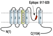
- Peptide SDVDGTTYDFAHC, corresponding to amino acid residues 917-929 of human TRPM8 (Accession Q7Z2W7). 3rd extracellular loop.

 Western blot analysis of prostate carcinoma cell lines DU145 (lanes 1 and 3), Human LNCaP prostate carcinoma (lanes 2 and 4) and mouse-TRPM8 transfected HEK-293 (lanes 5 and 6) cell lysates:1,2,5. Anti-TRPM8 (extracellular) Antibody (#ACC-049), (1:200).
3,4,6. Anti-TRPM8 (extracellular) Antibody, preincubated with TRPM8 (extracellular) Blocking Peptide (#BLP-CC049).
- Transfected HEK-293 cells (Zeevi, D.A. et al. (2010) J. Cell Sci. 123, 3112.).
 Expression of TRPM8 in rat DRGImmunohistochemical staining of rat dorsal root ganglion (DRG) frozen sections using Anti-TRPM8 (extracellular) Antibody (#ACC-049), (1:100). TRPM8 is expressed in DRG neurons. Hoechst 33342 is used as the counterstain.
 Multiplex staining of TRPM8 and TrkA in rat DRGImmunohistochemical staining of perfusion-fixed frozen rat dorsal root ganglia (DRG) sections using Anti-TrkA (extracellular)-ATTO Fluor-633 Antibody (#ANT-018-FR), (1:60) and Anti-TRPM8 (extracellular) Antibody (#ACC-049), (1:300). A. TRPM8 labeling followed by goat-anti-rabbit-Alexa-488 (green). B. The same section was then labeled for TrkA (purple). C. Merge of A and B demonstrates co-localization of TRPM8 and TrkA in rat DRG (arrows). Cell nuclei were stained with DAPI (blue).
 Expression of TRPM8 in rat DRG cellsImmunocytochemistry of rat dorsal root ganglion (DRG) cells. A. Intracellular staining of cells with Anti-TRPM8 (extracellular) Antibody (#ACC-049), (1:500) followed by goat anti-rabbit-AlexaFluor-555 secondary antibody. B. Extracellular staining of live cells with Anti-TRPM8 (extracellular) Antibody (1:50) followed by goat anti-rabbit-AlexaFluor-555 secondary antibody. The cell-permeable dye Hoechst 33342 (blue) was used for nuclear staining.
- Human prostate cancer DU145 cells (1:50) (Wang, Y. et al. (2012) Pathol. Oncol. Res. 18, 903.).
- Human, mouse and rat TRPM8 expressed in CHO cells, and rat DRGs (Miller, S. et al. (2014) PLoS ONE 9, e107151).
 Expression of TRPM8 in LNCaP prostate carcinoma cell lineCell surface detection of TRPM8 in LNCaP cells with Anti-TRPM8 (extracellular) Antibody (#ACC-049). A. Extracellular staining of intact LNCaP cells with Anti-TRPM8 (extracellular) Antibody (1:100), followed by goat anti-rabbit-AlexaFluor-550 (red), (x100). B. Intracellular staining of LNCaP cells with Anti-TRPM8 (extracellular) Antibody (1:1000), followed by goat anti-rabbit-AlexaFluor-488 (green), (x100).
- Fleig, A. et al. (2004) Trends. Pharmacol. Sci. 25, 633.
- Brauchi, S. et al. (2004) Proc. Natl. Acad. Sci. 101, 15494.
- McKemy, D.D. et al. (2002) Nature 416, 52.
- Peier, A.M. et al. (2002) Cell 108, 705.
- Dragoni, I. et al. (2006) J. Biol. Chem. 281, 37353.
- Zhang, L. et al. (2006) Endocr. Relat. Cancer 13, 27.
The mammalian melastatin-related transient receptor potential (TRPM) is a subfamily of the TRP family. The family was named after the first member, melastatin (TRPM1), whose gene was identified in metastatic and benign melanomas.1 The TRPM proteins share structural homology with other members of the TRP superfamily channels; six putative transmembrane domains, and cytoplasmic N-terminus and C-terminus. However, due to their long N-terminus and C-terminus they were also named the long TRP channel family.1
The TRPM family consists of eight members designated as TRPM1-8 that can be further divided into four pairs: TRPM1 and TRPM3; TRPM2 and TRPM8; TRPM4 and TRPM5; and TRPM6 and TRPM7.1
The TRPM8 channel is the cold and menthol receptor activated by either cold temperatures(~28°C) or menthol.2
TRPM8 is expressed in dorsal root ganglia (DRGs) where about 5%-10% of the small diameter DRG neurons express the channel.3,4 In DRGs, TRPM8 expressing neurons do not express the TRPV1 channel.5 Overexpression of TRPM8 was found in prostate cancer cells. However, the physiological and pathological roles that these cells play is still elusive.6 In prostate, it was suggested that TRPM8 might play a possible role in the progression of cancer to the metastatic stage.6
Application key:
Species reactivity key:
Anti-TRPM8 (extracellular) Antibody (#ACC-049) is a highly specific antibody directed against an epitope of the human protein. The antibody can be used in western blot, immunoprecipitation, immunohistochemistry, immunocytochemistry, and neutralization applications. It has been designed to recognize TRPM8 from human, rat, and mouse samples.

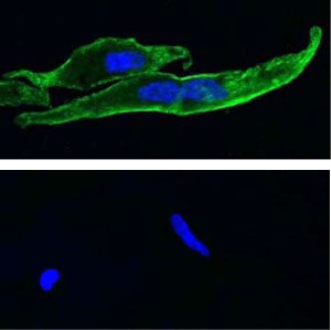
Expression of TRPM8 in rat tail artery astrocytes.Immunocytochemical staining of rat tail artery astrocytes using Anti-TRPM8 (extracellular) Antibody (#ACC-049). TRPM8 staining is shown in green, nuclei are in blue. Omission of the antibody (lower panel) abolishes TRPM8 staining.Adapted from Johnson, C.D. et al. (2009) Am. J. Physiol. 296, H1868. with permission of the American Physiological Society.
Applications
Citations
 Anti-TRPM8 (extracellular) Antibody specifically blocks TRPM8.A. Human TRPM8 cold activation is blocked by 2.5 μM of Anti-TRPM8 (extracellular) Antibody (#ACC-049). B. TRPM8 iciclin activation is blocked by 2.5 μM of the same antibody. M8-B is a specific TRPM8 antagonist. C. Anti-TRPM8 (extracellular) Antibody does not block cold activation of TRPA1 and heat activation of TRPV1 (D). AMG9090 and AMG6541 are specific TRPA1 and TRPV1 antagonist.
Adapted from Miller, S. et al. (2014) with kind permission of Dr. Gavva, N.R. Department of Neuroscience, Amgen Inc., California, U.S.A.
- Western blot analysis of TRPM8 transfected human HeLa cells. Tested in transfected cells treated with siRNA for TRPM8.
Huang, Y. et al. (2020) Front. Oncol. 10, 2645. - Western blot analysis and immunocytochemistry of human prostate cell lines. Tested in cells with TRPM8 knocked out using CRISPR-Cas9.
Alaimo, A. et al. (2020) Cell Death Dis. 11, 1039.
- Transfected HEK293 cell lysate (1:500).
Gkika, D. et al. (2015) J. Cell Biol. 208, 89. - Rat F11 dorsal root ganglion hybridoma cells lysate (1:400).
Toro, C.A. et al. (2015) J. Neurosci. 35, 571.
- Transfected HEK 293 cells (7 µg/ml).
Vinuela-Fernandez, I. et al. (2014) Neuropharmocolgy 79, 136. - Transfected HEK 293 cells.
Zeevi, D.A. et al. (2010) J. Cell Sci. 123, 3112.
- Rat DRG sections (1:300).
Vinuela-Fernandez, I. et al. (2014) Neuropharmocolgy 79, 136.
- Rat tail artery VSMC (1:200).
Melanaphy, D. et al. (2016) Am. J. Physiol. 311, H1416. - Rat F11 dorsal root ganglion hybridoma cells (1:400).
Toro, C.A. et al. (2015) J. Neurosci. 35, 571. - Human prostate cancer DU145 cells (1:50).
Wang, Y. et al. (2012) Pathol. Oncol. Res. 18, 903. - Rat tail artery astrocytes.
Johnson, C.D. et al. (2009) Am. J. Physiol. 296, H1868.
- Human, mouse and rat TRPM8 expressed in CHO cells, and rat DRGs.
Miller, S. et al. (2014) PLoS ONE 9, e107151.
- Ma, S. et al. (2012) J. Mol. Cell Biol. 4, 88.
- El Karim, I.A. et al. (2011) J. Endod. 37, 473.
- El Karim, I.A. et al. (2011) Pain 152, 2211.
- Harrington, A.M. et al. (2011) Pain 152, 1459.
- Martinez Lopez, P. et al. (2011) J. Cell Physiol. 226, 1620.
- Johnson, C.D. et al. (2009) Am. J. Physiol. 296, H1868.

