Overview
- Peptide (C)EDAEVFKDSMVPGEK, corresponding to amino acid residues 824-838 of rat TRPV1 (Accession O35433). Intracellular, C-terminus.

 Western blot analysis of human TRPV1 transfected HEK-293 cells:1. Anti-TRPV1 (VR1) Antibody (#ACC-030), (1:200).
2. Anti-TRPV1 (VR1) Antibody, preincubated with TRPV1/VR1 Blocking Peptide (#BLP-CC030). Western blot analysis of rat DRG lysate:1. Anti-TRPV1 (VR1) Antibody (#ACC-030), (1:200).
2. Anti-TRPV1 (VR1) Antibody, preincubated with TRPV1/VR1 Blocking Peptide (#BLP-CC030).
 Expression of TRPV1 in rat DRGImmunohistochemical staining of rat dorsal root ganglion (DRG) using Anti-TRPV1 (VR1) Antibody (#ACC-030). A. TRPV1 (red) in DRG neurons. B. Staining with mouse anti-Parvalbumin (green) in the same DRG section. C. Confocal merge of TRPV1 and Parvalbumin demonstrates colocalization (arrows).
 Expression of TRPV1 in rat DRG primary cultureImmunocytochemical staining of paraformaldehyde-fixed and permeabilized rat dorsal root ganglia (DRG) primary cells with Anti-TRPV1 (VR1) Antibody (#ACC-030), (1:200). A. Staining followed by goat anti-rabbit-AlexaFluor-555 secondary antibody. B. Nuclear staining using the cell-permeable dye Hoechst 33342. C. Merged image of panels A and B.
- Human sperm cells (De Toni, L. et al. (2016) PLoS ONE 11, e016722).
- Human sperm cells (De Toni, L. et al. (2016) PLoS ONE 11, e016722).
- Gunthorpe, M.J. et al. (2002) Trends Pharmacol. Sci. 23, 183.
- Cesare, P. et al. (1999) Proc. Natl. Acad. Sci. U.S.A. 96, 7658.
- Kim, J. et al. (2003) Nature 424, 81.
- Ahern, G. P. et al. (2003) J. Biol. Chem. 278, 30429.
- Ross, R.A. et al. (2003) Br. J. Pharmacol. 140, 790.
- Tominaga, M. et al. (1998) Neuron 21, 531.
- Vlachova, V. et al. (2003) J. Neurosci. 23, 1340.
- Agopyan, N. et al. (2004) Am. J. Physiol. 286, L563.
- Reilly, C.A. et al. (2003) Toxicol. Sci. 73, 170.
TRPV1 (also named VR1, capsaicin receptor and vanilloid receptor) is a member of the transient receptor potential (TRP) channel family, which includes TRPC, TRPM, TRPA, TRPP, TRPML and the TRPV subfamilies. The TRPV subfamily consists of six members named, TRPV1-6. The TRPV1 channel is a vanilloid gated, nonselective cation channel.
The channel has sequence homology to the TRP family, and shares a similar predicted structure of six transmembrane domain (TM) with a pore loop between TM5 and TM6.1 TRPV1 is expressed predominantly in nociceptors and in sensory neurons.2,3
TRPV1 has many activators among them heat, protons, vanilloids like capsaicin, resiniferatoxin (RTX), and lipids. This channel is associated with tissue injury and inflammation.5,6
Other members of this family, TRPV2, TRPV3 and TRPV4 also show thermal sensitivity.7 Conformational changes in the C-terminus are responsible for many functions such as permeation and gating.
There is also evidence that deletion of the C-terminus causes a loss of sensitivity to any stimuli.7 Recent studies demonstrated involvement of TRPV1 in apoptosis where inhibition of the receptor prevented apoptosis.8,9
Application key:
Species reactivity key:
Anti-TRPV1 (VR1) Antibody (#ACC-030) is a highly specific antibody directed against an epitope of the rat protein. The antibody can be used in western blot, immunohistochemistry, and immunocytochemistry applications. It has been designed to recognize TRPV1 from mouse, rat, and human samples.

Knockout validation of Anti-TRPV1 (VR1) Antibody in mouse adipose tissue lysate.Western blot analysis of mouse adipose tissue lysate using Anti-TRPV1 (VR1) Antibody (#ACC-030). TRPV1 is not detected in TRPV1-/- animals.Adapted from Chen, J. et al. (2015) Cardiovasc. Diabetol. 14, 22. with permission of BioMed Central.
Applications
Citations
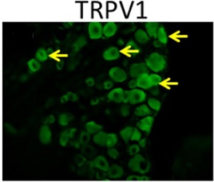
 Expression of TRPV1 in mouse DRGsImmunohistochemical staining of mouse DRG sections using Anti-TRPV1 (VR1) Antibody (#ACC-030). TRPV1 positive cells are detected in green.
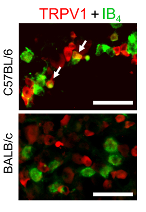
Adapted from Lin, J.G. et al. (2015) PLoS ONE 10, e0128037. with permission of PLoS. Differential expression of TRPV1 in C57BL/6 and BALB/c mice TG neurons.Immunohistochemical staining of trigeminal (TG) sections from C57BL/6 and BALB/c mice using Anti-TRPV1 (VR1) Antibody (#ACC-030). TRPV1 staining (green) is higher in IB4-positive TG neurons from C57BL/6 than BALB/c mouse strains.
Adapted from Ono, K. et al. (2015) with permission of American Physiological Society. TRPV1 co-localizes with CGRP in mouse DRG neurons.Immunocytochemical staining of mouse DRG neurons using Anti-TRPV1 (VR1) Antibody (#ACC-030). TRPV1 (green) co-localizes with CGRP (red) in large dense core vesicles (LDCVs).
Adapted from Devesa, I. et al. (2014) with permission of the National Academy of Sciences, USA.
- Western blot analysis of mouse adipose tissue lysate. Tested in TRPV1-/- mice.
Chen, J. et al. (2015) Cardiovasc. Diabetol. 14, 22.
- Mouse myoblasts (1:1000).
Obi, S. et al. (2017) J. Appl. Physiol. 122, 683. - Human sperm lysate.
De Toni, L. et al. (2016) PLoS ONE 11, e016722. - Mouse DRG lysate (1:1000).
Khoutorsky, A. et al. (2016) Proc. Natl. Acad. Sci. U.S.A. 113, 11949. - Human placenta lysate (1:500).
Martinez, N. et al. (2016) Placenta 40, 25. - Mouse adipose tissue lysate. Also tested in TRPV1-/- mice.
Chen, J. et al. (2015) Cardiovasc. Diabetol. 14, 22. - Rat brain lysate.
Nam, J.H. et al. (2015) Brain 138, 3610. - Human brain lysate.
Nam, J.H. et al. (2015) Brain 138, 3610. - Mouse brain stem and spinal cord lysates (1:500).
Kim, H.S. et al. (2014) Carcinogenesis 35, 1652. - Human peripheral blood mononuclear cells (PBMCs) and Natural Killer cells (NK) (1:500).
Kim, H.S. et al. (2014) Carcinogenesis 35, 1652.
- Rat DRG sections (1:500).
Morgan, M. et al. (2019) Bone 123, 168. - Human testis sections.
De Toni, L. et al. (2016) PLoS ONE 11, e016722. - Rat DRG sections (1:100).
Doppler, K. et al. (2016) Brain 139, 2617. - Human placenta sections (1:100).
Martinez, N. et al. (2016) Placenta 40, 25. - Mouse DRG sections.
Lin, J.G. et al. (2015) PLoS ONE 10, e0128037. - Rat brain sections.
Nam, J.H. et al. (2015) Brain 138, 3610. - Human brain sections.
Nam, J.H. et al. (2015) Brain 138, 3610. - Mouse trigeminal sections (1:400).
Ono, K. et al. (2015) J. Neurophysiol. 113, 3345. - Rat trigeminal sections (1:500).
Urata, K. et al. (2015) J. Dent. Res. 94, 446. - Human uveal melanoma sections (1:200).
Mergler, S. et al. (2014) Cell. Signal. 26, 56. - Rat DRG sections (1:1000).
Isensee, J. et al. (2014) J. Cell Sci. 127, 216. - Human skin sections (1:1000).
Cheng, H.T. et al. (2013) J. Pain. 14, 941.
- Mouse myoblasts.
Obi, S. et al. (2017) J. Appl. Physiol. 122, 683. - Human sperm cells.
De Toni, L. et al. (2016) PLoS ONE 11, e016722. - Mouse DRG neurons.
Devesa, I. et al. (2014) Proc. Natl. Acad. Sci. U.S.A. 111, 18345.
- Human sperm cells.
De Toni, L. et al. (2016) PLoS ONE 11, e016722.
- Human sperm cells.
De Toni, L. et al. (2016) PLoS ONE 11, e016722.
- Tanaka, Y. et al. (2016) Neuron 90, 1215.
- Belghiti, M. et al. (2013) J. Biol. Chem. 288, 9675.
- Efendiev, R. et al. (2013) J. Biol. Chem. 288, 3929.
- Jankowski, M.P. et al. (2013) J. Neurophysiol. 109, 2374.
- Lotteau, S. et al. (2013) PLoS ONE 8, e58673.
- Matsuura, S. et al. (2013) PLoS ONE 8, e52840.
- Jankowski, M.P. et al. (2012) J. Neurosci. 32, 17869.
