Overview
- Peptide CDGHQQGYAPKWRAEDAPL, corresponding to amino acid residues 853-871 of rat TRPV4 (Accession Q9ERZ8). Intracellular, C-terminus.
- Rat brain lysates and ND7/23 Mouse neuroblastoma /rat dorsal root ganglion neurone hybrid cell line lysate (1:200).
 Western blot analysis of rat brain lysates:1. Anti-TRPV4 Antibody (#ACC-034), (1:200).
2. Anti-TRPV4 Antibody, preincubated with TRPV4 Blocking Peptide (#BLP-CC034). Western blot analysis of ND7/23 Mouse neuroblastoma /rat dorsal root ganglion neuron hybrid cell line lysate:1. Anti-TRPV4 Antibody (#ACC-034), (1:200).
2. Anti-TRPV4 Antibody, preincubated with TRPV4 Blocking Peptide (#BLP-CC034).
- Rat and mouse brain lysates (10 μg Ab/0.500 mg whole-protein) (Benfenati, V. et al. (2011) Proc. Natl. Acad. Sci. U.S.A. 108, 2563).
- Rat brain and mouse cerebellum frozen sections.
- Rat brain and mouse cerebellum frozen sections.
- Human corneal epithelial cells (HCEC) (1:200) (Pan, Z. et al. (2008) Cell Calcium 44, 374.).
- The control antigen is not suitable for this application.
TRP channels are a large family (about 28 genes) of plasma membrane, non-selective cationic channels that are either specifically or ubiquitously expressed in excitable and non-excitable cells.1 According to IUPHAR the TRP family comprises of three main subfamilies on the basis of sequence homology; TRPC, TRPM and TRPV (to date, three extra subfamilies are considered to belong to the TRP family; the TRPA, TRPML, and TRPP).1-4 The TRPV subfamily consists of six members, TRPV1-6.5
TRPV4 (also named OTRPC4) is activated under hypotonic conditions and serves as an osmoreceptor. TRPV4 is expressed in brain, liver, kidney, heart, testis and salivary gland.6
Application key:
Species reactivity key:

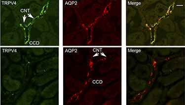
Knockout validation of Anti-TRPV4 Antibody in mouse kidney.Immunohistochemical staining of mouse kidney sections using Anti-TRPV4 Antibody (#ACC-034) and Anti-Aquaporin 2-ATTO Fluor-550 Antibody (#AQP-002-AO). TRPV4 staining (green, upper panels) is detected in both the connecting tubules (CNT) and the collecting ducts (CCD). AQP2 immunostaining (red) is also observed in the CCD and CNT. TRPV4 staining is not observed in the sections from TRPV4-/- mice. Adapted from Berrout, J. et al. (2012) J. Biol. Chem. 287, 8782. with permission of American Society for Biochemistry and Molecular Biology.
