Overview
- Peptide (C)DAVRLREVQGKDGGE, corresponding to amino acid residues 495-509 of rat VAChT (Accession Q62666). Cytoplasmic, C-terminus.
- Mouse and rat brain lysates.
 Western blot analysis of mouse (lanes 1 and 3) and rat (lanes 2 and 4) brain lysates:1,2. Anti-Vesicular Acetylcholine Transporter (VAChT) Antibody (#ACT-003), (1:200).
3,4. Anti-Vesicular Acetylcholine Transporter (VAChT) Antibody, preincubated with Vesicular Acetylcholine Transporter/VACHT Blocking Peptide (#BLP-CT003).
- Rat brain and spinal cord sections (1:400).
In cholinergic neurons, acetylcholine is synthesized from choline and acetyl-coenzyme A (acetyl-CoA). Vesicular acetylcholine transporter (VAChT) transports acetylcholine from the cytoplasm to the lumen of synaptic vesicles where following depolarization, acetylcholine is released in the synaptic cleft and activates muscarinic and nicotinic receptors located on postsynaptic membranes1-3. Acetylcholine released in the synaptic cleft is rapidly hydrolyzed into choline and acetate. Since choline is not de novo synthesized and thereby only made available through diet uptake, choline is recycled back into the cells in order to regenerate acetylcholine1,2.
VAChT is a member of the SLC18 family of transporters. It has 12 membrane spanning domains, with extracellular N- and C-terminal tails. The transporter exchanges cytoplasmic acetylcholine for two vesicular protons4-6.
VAChT is a selective marker of cholinergic neurons and could be used therefore to determine the loss of cholinergic neurons in Alzheimer’s or Alzheimer’s related disease7. VAChT knockout mice die after birth. They display a lack of stimulated release of acetylcholine and an underdeveloped neuromuscular junction4.
Application key:
Species reactivity key:
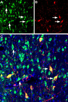
 Multiplex staining of Vesicular Acetylcholine Transporter and p75NTR in rat medial septum.Immunohistochemical staining of immersion-fixed, free floating rat brain frozen sections using rabbit Anti-Vesicular Acetylcholine Transporter (VAChT) Antibody (#ACT-003), (1:200) and Mouse Anti-Rat p75 NGF Receptor (extracellular) Antibody (#AN-170), (1:200). A. VAChT staining (green) appears in several neuronal cells. B. p75NTR (red) also stains neuronal cells. C. Merge of the two images shows colocalization of VAChT and p75NTR in some cells (horizontal arrows), while other cells express only VAChT (vertical arrows). Cell nuclei are stained with DAPI (blue).
