Overview
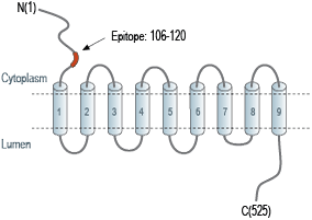
- Peptide (C)GEFGGHDKPKITAWE, corresponding to amino acid residues 106-120 of rat vesicular GABA transporter (Accession O35458). Cytoplasmic, N-terminus.

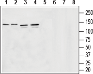
 Western blot analysis of rat brain membranes (lanes 1 and 5), mouse brain membranes (lanes 2 and 6), human U-87 MG glyoblastoma lysates (lanes 3 and 7) and human SH-SY5Y brain neuroblastoma lysates (lanes 4 and 8):1-4. Guinea pig Anti-Vesicular GABA Transporter (VGAT) Antibody (#AGT-005-GP), (1:200).
5-8. Guinea pig Anti-Vesicular GABA Transporter (VGAT) Antibody, preincubated with Vesicular GABA Transporter/VGAT Blocking Peptide (#BLP-GT005).
 Expression of VGAT in mouse cerebellum.Immunohistochemical staining of perfusion-fixed frozen mouse brain sections with Guinea pig Anti-Vesicular GABA Transporter (VGAT) Antibody (#AGT-005-GP), (1:600), followed by goat anti-guinea pig-AlexaFluor-594. A. VGAT immunoreactivity (red) appears in the granule layer (GL) and in “pinceau” structures adjacent to purkinje cells (arrows). B. Pre-incubation of the antibody with Vesicular GABA Transporter/VGAT Blocking Peptide (#BLP-GT005), suppressed staining. Cell nuclei are stained with DAPI (blue).
 Expression of VGAT in rat cerebellum.Immunohistochemical staining of perfusion-fixed frozen rat brain sections with Guinea pig Anti-Vesicular GABA Transporter (VGAT) Antibody (#AGT-005-GP), (1:600), followed by goat anti-guinea pig-AlexaFluor-594. A. VGAT immunoreactivity (red) appears in the granule layer (GL) and in “pinceau” structures adjacent to purkinje cells (arrows). B. Pre-incubation of the antibody with Vesicular GABA Transporter/VGAT Blocking Peptide (#BLP-GT005), suppressed staining. Cell nuclei are stained with DAPI (blue).
- Gasnier, B. (2004) Pflugers. Arch. 447, 756.
- McIntire, S. et al. (1997) Nature. 389, 870.
- Bedet, C. et al. (2000) J. Neurochem. 75, 1654.
- Chaudry, F. et al. (1998) J. Neurosci. 18, 9733.
- Dumoulin, A. et al. (1999) J. Cell. Sci. 112, 811.
- Takamori, S. et al. (2000) J. Neurosci. 20, 4904.
- Mayerhofer, A. et al. (2001) FASEB J. 15, 1089.
- Chessler, S. et al. (2002) Diabetes. 51, 1763.
The superfamily of amino acid transporters includes SLC32, SLC36 and SLC38 families. SLC32 has only one member, the vesicular inhibitory amino acid transporter (VIAAT) or vesicular GABA transporter (VGAT)1.
VGAT is responsible for the uptake of GABA and glycine in exchange of protons, and their release from nerve terminals1.
The transporter is organized into ten membrane spanning domains, a cytosolic N-terminus and a luminal C-terminal tail2. In many regions in the brain, where it is expressed, it is found constitutively phosphorylated, although the biological significance is currently unknown3.
VGAT is expressed on synaptic vesicles of GABAergic and glycynergic neurons4-6. The transporter is also detected in the pituitary gland and in the pancreas7,8.
Application key:
Species reactivity key:
Guinea pig Anti-Vesicular GABA Transporter (VGAT) Antibody (#AGT-005-GP, formerly AGP-129), raised in guinea pigs, is a highly specific antibody directed against an epitope of the rat protein. The antibody can be used in western blot analysis. It has been designed to recognize VGAT from mouse, rat, and human samples. The antigen used to immunize guinea pigs is the same as Anti-Vesicular GABA Transporter (VGAT) Antibody (#AGT-005) raised in rabbit. Our line of guinea pig antibodies enables more flexibility with our products such as multiplex staining studies, immunoprecipitation, etc.
