Overview
Cat #: BLP-CC003
Type: Synthetic peptide
Form: Lyophilized powder
Cav1.2/CACNA1C Blocking Peptide (#BLP-CC003) is the original antigen used for immunization during Anti-CaV1.2 (CACNA1C) Antibody (#ACC-003) generation. The blocking peptide binds and ‘blocks’ Anti-Cav1.2/CACNA1C primary antibody, this makes it a good negative reagent control to help confirm antibody specificity in western blot and immunohistochemistry applications. This control is also often called a pre-adsorption control.
Applications: wb, ihc
Application key:
WB- Western blot, IHC- Immunohistochemistry
For research purposes only. not for human use
Applications
Demonstration of Pre-adsorption control
 Western blot analysis of rat brain membranes:1. Anti-CaV1.2 (CACNA1C) Antibody (#ACC-003), (1:200).
2. Anti-CaV1.2 (CACNA1C) Antibody, preincubated with Cav1.2/CACNA1C Blocking Peptide (#BLP-CC003). Western blot analysis of CaV1.2-transfected Xenopus oocytes (lane 1) and non-transfected oocytes lysates (lane 2):1. Anti-CaV1.2 (CACNA1C) Antibody (#ACC-003), (1:200) in CaV1.2 (CACNA1C) Channel Overexpressed in Xenopus oocytes.
2. Anti-CaV1.2 (CACNA1C) Antibody in non-transfected oocytes. Expression of CaV1.2 in mouse cerebellumImmunohistochemical staining of mouse cerebellum with Anti-CaV1.2 (CACNA1C) Antibody (#ACC-003). A. CaV1.2 (red) appears in Purkinje cells (horizontal arrows) and is distributed diffusely in the molecular layer (Mol) including in Purkinje dendrites (vertical arrows). B. Staining of Purkinje nerve cells with mouse anti-Calbindin 28K (green) demonstrates the location of dendrites in the molecular layer. C. Merged image of panels A and B.
- Mouse atrioles (1:300) (Howitt, L. et al. (2013) J. Physiol. 591, 2157.).
 Western blot analysis of rat brain membrane:1. Guinea pig Anti-CaV1.2 (CACNA1C) Antibody (#ACC-003-GP), (1:200).
2. Guinea pig Anti-CaV1.2 (CACNA1C) Antibody, preincubated with Cav1.2/CACNA1C Blocking Peptide (#BLP-CC003). Western blot analysis of CaV1.2-transfected Xenopus oocytes (lane 1) and non-transfected oocytes lysates (lane 2):1. Guinea pig Anti-CaV1.2 (CACNA1C) Antibody (#ACC-003-GP), (1:200) in CaV1.2 (CACNA1C) Channel Overexpressed in Xenopus oocytes.
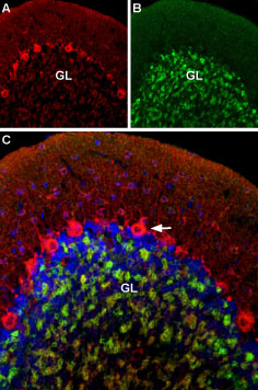
2. Guinea pig Anti-CaV1.2 (CACNA1C) Antibody in non-transfected oocytes. Multiplex staining of CaV1.2 and GABA(A) α1 Receptor in rat cerebellumImmunohistochemical staining of rat cerebellum using Guinea pig Anti-CaV1.2 (CACNA1C) Antibody (#ACC-003-GP) and Anti-GABA(A) α1 Receptor (extracellular)-ATTO Fluor-488 Antibody (#AGA-001-AG). A. CaV1.2 (red) is detected mostly in Purkinje cells (arrow). B. In the same section, GABA(A) α1 Receptor (green) is observed in the granule layer. C. Merge of the two images suggests some colocalization between CaV1.2 and GABA(A) α1 Receptor in the rat granule layer but only CaV1.2 appears in Purkinje cells.
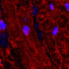
 Expression of CaV1.2 in human atriaImmunohistochemical staining of human left atrium using Guinea pig Anti-CaV1.2 (CACNA1C) Antibody (#ACC-003-GP), (1:100).
The picture was kindly provided by Dr. Van Wagoner, D.R. from the Department of Molecular Cardiology, Cleveland Clinic, Cleveland, Ohio, USA. Lovano, B. and Peterson, J. collected the data. Expression of CaV1.2 in rat heartImmunohistochemical staining of rat heart paraffin embedded sections using Guinea pig Anti-CaV1.2 (CACNA1C) Antibody (#ACC-003-GP). A. CaV1.2 staining (green) appears mainly in the cardiac muscle, and in a lesser intensity in the tunica intima layer of the smooth muscle of the muscular arteries. B. Nuclear staining using DAPI as the counter stain. C. Merged images of A and B.
 Expression of CaV1.2 in mouse hippocampusImmunohistochemical staining of mouse dentate gyrus using Guinea pig Anti-CaV1.2 (CACNA1C) Antibody (#ACC-003-GP). A. CaV1.2 (green) appeared in the outer molecular layer of the dentate gyrus and in the granule layer. B. Counterstain with DAPI (blue) outlines the granule layer of the dentate gyrus.
Properties
Sequence
Accession (Uniprot) Number P22002
Peptide Confirmation Confirmed by amino acid analysis and mass spectrometry.
Purity >70%
Storage Before Reconstitution Lyophilized powder can be stored intact at room temperature for two weeks. For longer periods, it should be stored at -20°C.
Reconstitution 100 µl double distilled water (DDW).
Concentration After Reconstitution 0.4 mg/ml.
Storage After Reconstitution -20°C.
Antigen Preadsorption Control 1 µg peptide per 1 µg antibody.
Standard Quality Control Of Each Lot Western blot analysis.

