Overview
Type: Synthetic peptide
Form: Lyophilized powder
Connexin-43 Blocking Peptide (#BLP-CC201) is the original antigen used for immunization during Anti-Connexin-43 Antibody (#ACC-201) generation. The blocking peptide binds and ‘blocks’ Anti-Connexin-43 primary antibody, this makes it a good negative reagent control to help confirm antibody specificity in western blot and immunohistochemistry applications. This control is also often called a pre-adsorption control.
Applications: wb, ihc
For research purposes only. not for human use
Applications
Demonstration of Pre-adsorption control
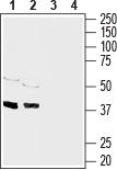
 Western blot analysis of mouse brain membranes:1. Anti-Connexin-43 Antibody (#ACC-201), (1:400).
2. Anti-Connexin-43 Antibody, preincubated with Connexin-43 Blocking Peptide (#BLP-CC201). Western blot analysis of rat heart membranes:1. Anti-Connexin-43 Antibody (#ACC-201), (1:400).
2. Anti-Connexin-43 Antibody, preincubated with Connexin-43 Blocking Peptide (#BLP-CC201). Expression of Connexin-43 in rat cerebellumImmunohistochemical staining of immersion-fixed, free floating rat brain frozen sections using Anti-Connexin-43 Antibody (#ACC-201), (1:300). Cx43 staining (red) appeared in Bergmann glial fibers (arrows) in the molecular layer (MOL) and in the granule layer (G). Cell nuclei were stained with DAPI (blue).
- Human fetal cochlear sections (Locher, H. et al. (2015) Dev. Neurobiol. 75, 1219.).
 Western blot analysis of rat brain (lanes 1 and 3), (1:1000) and mouse brain (lanes 2 and 4), (1:200) membranes:1,2. Guinea pig Anti-Connexin-43 Antibody (#ACC-201-GP).
3,4. Guinea pig Anti-Connexin-43 Antibody, preincubated with Connexin-43 Blocking Peptide (#BLP-CC201). Western blot analysis of rat (lanes 5 and 7) and mouse (lanes 6 and 8) heart membranes:5,6. Guinea pig Anti-Connexin-43 Antibody (#ACC-201-GP), (1:400).
7,8. Guinea pig Anti-Connexin-43 Antibody, preincubated with Connexin-43 Blocking Peptide (#BLP-CC201).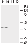 Western blot analysis of human T-cell leukemia (Jurkat) (lanes 9 and 11) and human neuroblastoma (SH-SY5Y) (lanes 10 and 12) cell line lysates:9,10. Guinea pig Anti-Connexin-43 Antibody (#ACC-201-GP), (1:400).
11,12. Guinea pig Anti-Connexin-43 Antibody, preincubated with Connexin-43 Blocking Peptide (#BLP-CC201). Expression of Connexin-43 in mouse brainImmunohistochemical staining of frozen mouse cerebellum free floating sections using Guinea pig Anti-Connexin-43 Antibody (#ACC-201-GP). CX43 immunoreactivity (red) appears in the Purkinje layer (vertical arrows) and in puncta along dendritic processes (horizontal arrows). Nuclei are stained using DAPI as the counterstain (blue).
 Multiplex staining of Connexin-43 and GFAP in rat cerebellum.Immunohistochemical staining of perfusion-fixed frozen rat brain sections with Guinea pig Anti-Connexin-43 Antibody (#ACC-201-GP), (1:200), followed by goat anti-guinea pig-AlexaFluor-594 and Anti-GFAP Antibody (#AFP-001), (1:1200), followed by goat anti-rabbit-AlexaFluor-488. A. Connexin-43 immunoreactivity (red) appears as positive puncta (arrows). B. GFAP immunoreactivity (green) appears along the Bergmann glial processes (arrows point at examples). C. Merge of the two images shows CNX-43 puncta distributed along the Bergmann glial processes. Cell nuclei are stained with DAPI (blue).
Properties
Sequence
- (C)HAQPFDFPDDNQNSK, corresponding to amino acids residues 331-345 of human Connexin-43 (Accession P17302).

Accession (Uniprot) Number P17302
Peptide Confirmation Confirmed by amino acid analysis and mass spectrometry.
Purity >70%
Storage Before Reconstitution Lyophilized powder can be stored intact at room temperature for two weeks. For longer periods, it should be stored at -20°C.
Reconstitution 100 µl double distilled water (DDW).
Concentration After Reconstitution 0.4 mg/ml.
Storage After Reconstitution -20°C.
Antigen Preadsorption Control 1 µg peptide per 1 µg antibody.
Standard Quality Control Of Each Lot Western blot analysis.
