Overview
Cat #: BLP-PC006
Type: GST fusion protein
Form: Lyophilized powder
GIRK2/Kir3.2 Blocking Peptide (#BLP-PC006) is the original antigen used for immunization during Anti-GIRK2 (Kir3.2) Antibody (#APC-006) generation. The blocking peptide binds and ‘blocks’ Anti-GIRK2/Kir3.2 primary antibody, this makes it a good negative reagent control to help confirm antibody specificity in western blot and immunohistochemistry applications. This control is also often called a pre-adsorption control.
Applications: wb, ihc
Application key:
WB- Western blot, IHC- Immunohistochemistry
For research purposes only. not for human use
Applications
Demonstration of Pre-adsorption control
 Western blot analysis of rat brain membranes:1. Anti-GIRK2 (Kir3.2) Antibody (#APC-006), (1:200).
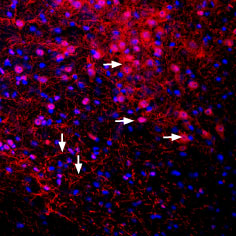
2. Anti-GIRK2 (Kir3.2) Antibody, preincubated with GIRK2 (Kir3.2) Blocking peptide (#BLP-PC006). Expression of Kir3.2 in mouse brainImmunohistochemical staining of perfusion-fixed frozen mouse substantia nigra pars compacta sections using Anti-GIRK2 (Kir3.2) Antibody (#APC-006), (1:400). GIRK2 staining (red) appears in cells and processes along the pars compacta of the mouse substantia nigra (horizontal arrows) and in the pars reticulata (vertical arrows). Cell nuclei are stained with DAPI (blue).
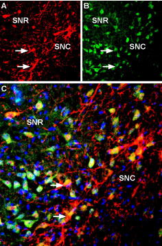
 Multiplex staining of Kir3.2 and KCNK2 in rat substantia nigraImmunohistochemical staining of immersion-fixed, free floating rat brain frozen sections using rabbit Anti-GIRK2 (Kir3.2) Antibody (#APC-006), (1:400) and Guinea pig Anti-KCNK2 (TREK-1) Antibody (#APC-047-GP), (1:120). A. Kir3.2 staining (red) appears in cells of the substantia nigra pars compacta (SNC, arrows). B. KCNK2 (green) appears in both compacta (SNC) and reticulata (SNR) portions of the substantia nigra. C. Merge of the two images reveals co-localization in some cells (arrows), mainly in the SNC region. Cell nuclei are stained with DAPI (blue).
- Mouse spinal cords (Marker, C.L. et al. (2004) J. Neurosci. 24, 2806.).
 Western blot analysis of rat brain membrane (lanes 1 and 3) and mouse brain membrane (lanes 2 and 4):1,2. Guinea pig Anti-GIRK2 (Kir3.2) Antibody (#APC-006-GP), (1:400).
3,4. Guinea pig Anti-GIRK2 (Kir3.2) Antibody, preincubated with GIRK2/Kir3.2 Blocking Peptide (#BLP-PC006).
Following a screen of several secondary antibodies, the following were used for western blot analysis:
Anti-Guinea pig IgG (Sigma #A7289 or Jackson ImmunoResearch #106-035-006). Multiplex staining of GIRK2 (Kir3.2) and LRRK2 in mouse substantia nigra pars compactaImmunohistochemical staining of immersion-fixed, free floating mouse brain frozen sections using Guinea pig Anti-GIRK2 (Kir3.2) Antibody (#APC-006-GP), (1:200) and rabbit Anti-LRRK2 Antibody (#ANR-102), (1:200). A. Kir3.2 staining (red) appears in profiles of dopaminergic neurons (arrows). B. LRRK2 staining (green) is detected in several cell bodies in the SNC. C. Merge of the two images demonstrates colocalization in several neurons (arrows). Cell nuclei are stained with DAPI (blue).
 Expression of GIRK2 (Kir3.2) in mouse brainImmunohistochemical staining of mouse substantia nigra pars compacta (SNC) using Guinea pig Anti-GIRK2 (Kir3.2) Antibody (#APC-006-GP), (1:400). A. GIRK2 staining (green) appears in soma (horizontal arrows) and dendrites of dopaminergic neurons (vertical arrow). B. Nuclei staining using DAPI as the counterstain (blue). C. Merged image of panels A and B.
 Expression of GIRK2 (Kir3.2) in rat brainImmunohistochemical staining of rat substantia nigra pars compacta (SNC) using Guinea pig Anti-GIRK2 (Kir3.2) Antibody (#APC-006-GP), (1:400). A. GIRK2 staining (green) appears in soma (horizontal arrows) and dendrites of dopaminergic neurons (vertical arrow). B. Nuclei staining using DAPI as the counterstain (blue). C. Merged image of panels A and B.
Properties
Sequence
Accession (Uniprot) Number P48542
Peptide Confirmation Confirmed by DNA sequence and SDSPAGE.
Purity >95% (SDS-PAGE)
Storage Before Reconstitution Lyophilized powder can be stored intact at room temperature for two weeks. For longer periods, it should be stored at -20°C.
Reconstitution 100 μl PBS.
Concentration After Reconstitution 1.2 mg/ml.
Storage After Reconstitution -20°C.
Antigen Preadsorption Control 3 µg fusion protein per 1 µg antibody.
Standard Quality Control Of Each Lot Western blot analysis.

