Overview
Type: Synthetic peptide
Form: Lyophilized powder
GluR1/GluA1 (extracellular) Blocking Peptide (#BLP-GC004) is the original antigen used for immunization during Anti-GluR1 (GluA1) (extracellular) Antibody (#AGC-004) generation. The blocking peptide binds and ‘blocks’ Anti-GluR1/GluA1 (extracellular) primary antibody, this makes it a good negative reagent control to help confirm antibody specificity in western blot and immunohistochemistry applications. This control is also often called a pre-adsorption control.
Applications: wb, ihc
For research purposes only. not for human use
Applications
Demonstration of Pre-adsorption control
 Western blot analysis of rat (lanes 1 and 3) and mouse (lanes 2 and 4) brain lysates:1,2. Anti-GluR1 (GluA1) (extracellular) Antibody (#AGC-004), (1:200).
3,4. Anti-GluR1 (GluA1) (extracellular) Antibody, preincubated with GluR1/GluA1 (extracellular) Blocking Peptide (#BLP-GC004).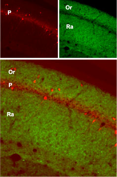 Expression of GluR1 in mouse hippocampusImmunohistochemical staining of mouse hippocampus with Anti-GluR1 (GluA1) (extracellular) Antibody (#AGC-004). GluR1 (green) is present in the stratum oriens (Or) and radiatum (Ra) but not in the pyramidal layer (P). Staining of the same section with mouse anti-parvalbumin (red) identifies the pyramidal layer.
 Western blot analysis of rat (lanes 1 and 3) and mouse (lanes 2 and 4) brain lysates:1,2. Guinea pig Anti-GluR1 (GluA1) (extracellular) Antibody (#AGC-004-GP), (1:200).
3,4. Guinea pig Anti-GluR1 (GluA1) (extracellular) Antibody, preincubated with GluR1/GluA1 (extracellular) Blocking Peptide (#BLP-GC004). Expression of GluR1 in rat hippocampusImmunohistochemical staining of perfusion-fixed frozen rat brain sections using Guinea pig Anti-GluR1 (GluA1) (extracellular) Antibody (#AGC-004-GP), (1:400), followed by goat anti-guinea pig-Cy2 antibody (green). GluR1 staining appears in neuronal outlines (horizontal arrows) and in the inner molecular layer of the dentate gyrus (vertical arrow). Nuclei are stained with DAPI (blue).
 Multiplex staining of GluR1 and PICK1 in rat hippocampus.Immunohistochemical staining of perfusion-fixed frozen rat brain sections with Guinea pig Anti-GluR1 (GluA1) (extracellular) Antibody (#AGC-004-GP), (1:400), followed by goat anti-guinea pig-AlexaFluor-488 and Anti-PICK1 Antibody (#APZ-014), (1:400), followed by donkey anti-rabbit-Cy3. A. GluR1 immunoreactivity (green) appears in several interneurons (arrows). B. PICK1 immunoreactivity (red) appears also in interneurons (arrows). C. Merge of the two images reveals co-localization in several interneurons (arrows) in the dentate gyrus (DG). Cell nuclei are stained with DAPI (blue).
Properties
Sequence
- RTSDSRDHTRVDWKR(C), corresponding to amino acid residues 271-285 of rat GluR1 (Accession P19490).

Accession (Uniprot) Number P19490
Peptide Confirmation Confirmed by amino acid analysis and mass spectrometry.
Purity >70%
Storage Before Reconstitution Lyophilized powder can be stored intact at room temperature for two weeks. For longer periods, it should be stored at -20°C.
Reconstitution 100 µl double distilled water (DDW).
Storage After Reconstitution -20°C.
Antigen Preadsorption Control 1 µg peptide per 1 µg antibody.
Standard Quality Control Of Each Lot Western blot analysis.
