Overview
- Ostrow, K.L. et al. (2003) Toxicon 42, 263.
- Suchyna, T.M. et al. (2000) J. Gen. Physiol. 115, 583.
- Redaelli, E. et al. (2010) J. Biol. Chem. 285, 4130.
 Alomone Labs GsMTx-4 inhibits NaV1.7 channel currents expressed in Xenopus oocytes.NaV1.7 currents were elicited by 100 ms voltage ramp from a holding potential of -100 mV to +30 mV, applied every 10 sec using whole-cell voltage clamp technique. Left: Superimposed traces of NaV1.7 currents before (black) and during (green) application of 500 nM GsMTx-4 (#STG-100). Right: GsMTx-4 dose response inhibition of NaV1.7 currents.
GsMTx-4 is a 34 amino acid peptidyl toxin originally isolated from the Grammostola rosea (Chilean rose) tarantula venom and belongs to the huwentoxin-1 family1.
This toxin inhibits different channels and in addition has antimicrobial activity. It blocks cation-selective mechanosensitive ion channels (strech-activated channels, SACs), without having an effect on whole-cell voltage-sensitive currents1. In addition, it inhibits atrial fibrillation2 as well as the membrane motor of outer hair cells3 at low doses. A medium toxicity on a large spectra of voltage-gated Na+ channels, namely NaV1.1, NaV1.2, NaV1.3, NaV1.4, NaV1.5, NaV1.6 and NaV1.7 was reported. GsMTx-4 also inhibits K+ channels KV11.1 and KV11.2, whereas it does not inhibit K+ channels KV1.1 (IC50 > 85 µM), KV1.4 (IC50 > 85 µM) and KV11.3 (IC50 = 53 µM)4. GsMTx-4 was also found to inhibit both TRPC1 and TRPC6 channels5,6, as well as Piezo1, the mechanosensitive channel7.
Antimicrobial activity is shown against several Gram-positive and Gram-negative bacteria as well8.

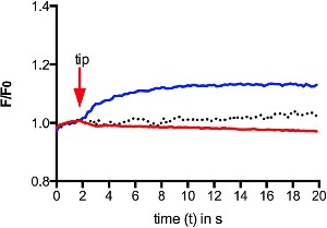
Alomone Labs GsMTx-4 inhibits mechanical-induced Ca2+ increase in human RBCs.Intracellular Ca2+ levels from Fluo-4 AM loaded human red blood cells (RBCs) were measured following mechanical stimulation touching a cell with a micropipette (blue). Application of 2.5 µM GsMTx-4 (#STG-100) prior to the stimulation blocked the intracellular Ca2+ increase (red). Control, without stimulation is depicted by the dotted graph.Adapted from Danielczok, J.G. et al. (2017) Front. Physiol. 8, 979. with permission of Frontiers.

