Overview
- Peptide (C)KFAKDDSDSMSRR, corresponding to amino acid residues 378-390 of rat AVPR1A (Accession P30560). Intracellular, C-terminus.

Vasopressin V1A Receptor/AVPR1A Blocking Peptide (BLP-VR010)
 Western blot analysis of rat brain membranes (lanes 1 and 3) and mouse brain membranes (lanes 2 and 4):1, 2. Guinea Pig Anti-Vasopressin V1A Receptor (AVPR1A) Antibody (#AVR-010-GP), (1:200).
3, 4. Guinea Pig Anti-Vasopressin V1A Receptor (AVPR1A) Antibody, preincubated with Vasopressin V1A Receptor (AVPR1A) Blocking Peptide (#BLP-VR010). Western blot analysis of rat kidney membranes (lanes 1 and 3) and mouse kidney membranes (lanes 2 and 4):1, 2. Guinea Pig Anti-Vasopressin V1A Receptor (AVPR1A) Antibody (#AVR-010-GP), (1:200).
3, 4. Guinea Pig Anti-Vasopressin V1A Receptor (AVPR1A) Antibody, preincubated with Vasopressin V1A Receptor (AVPR1A) Blocking Peptide (#BLP-VR010).
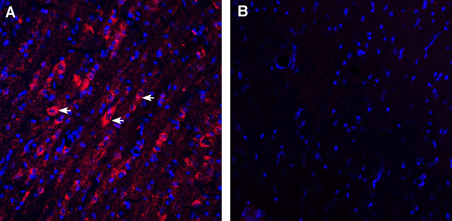
 Expression of V1aR in rat medial septum.Immunohistochemical staining of perfusion-fixed frozen rat brain sections with Guinea Pig Anti-Vasopressin V1A Receptor (AVPR1A) Antibody (#AVR-010-GP), (1:200), followed by goat anti-guinea pig - Alexa Fluor-594. A. V1aR immunoreactivity (red) appears in neuronal outlines (arrows). B. Pre-incubation of the antibody with Vasopressin V1A Receptor (AVPR1A) Blocking Peptide (#BLP-VR010), suppressed staining. Cell nuclei are stained with DAPI (blue).
- Thibonnier, M. et al. (1994) J. Biol. Chem. 269, 3304.
- Michell, R.H. et al. (1979) Biochem. Soc. Trans. 7, 861.
- Eisenberg, D. et al. (1984) Proc. Natl. Acad. Sci. U.S.A. 81, 140.
- Thibonnier, M. (1993) in neuroendocrinology of the concepts in Neurosurgery Series 5 (Selman, W., ed) pp. 19, Williams & Wilkins, Baltimore.
Vasopressin (AVP), the antidiuretic hormone, is a cyclic nonapeptide involved in the homeostasis of body fluid, blood volume, vascular tone, and blood pressure. AVP also belongs to the family of vasoactive and mitogenic peptides involved in physiological and pathological cell growth and differentiation1.
AVP exerts its actions through binding to specific V1A, V1B, and V2, membrane receptors coupled to distinct second messengers2. V1 AVP receptor has the typical features of a G-protein coupled transmembrane receptor with seven putative hydrophobic domains, connected by three extracellular and three intracellular loops3.
V1 receptors activate phospholipases A2, C, and D, resulting in the production of inositol 1,4,5-trisphosphate (IPS) and 1,Z-diacylglycerol (DAG), the mobilization of intracellular Ca2+, the influx of extracellular Ca2+, the activation of protein kinase C, and protein phosphorylation4. V1A AVP receptors have been shown by radioligand binding techniques to be present in vascular smooth muscle cells, hepatocytes, blood platelets, lymphocytes and monocytes, type 2 pneumocytes, adrenal cortex, brain (hippocampus septum et amygdalae), reproductive organs, retinal epithelium, renal mesangial cells, and the A10, A7r5,3T3, and WRK-1 cell lines4. V1A AVP receptors mediate cell contraction and proliferation, platelet aggregation, coagulation factor release, and glycogenolysis. V1B AVP receptors are located in the anterior pituitary where they stimulate ACTH release1.
Application key:
Species reactivity key:
Guinea Pig Anti-Vasopressin V1A Receptor (AVPR1A) Antibody (#AVR-010-GP) is a highly specific antibody directed against an epitope of the rat protein. The antibody can be used in western blot and immunohistochemistry applications. It has been designed to recognize V1aR from mouse, rat and human samples.
The antigen used to immunize guinea pigs is the same as Anti-Vasopressin V1A Receptor (AVPR1A) Antibody (#AVR-010) raised in rabbit. Our line of guinea pig antibodies enables more flexibility with our products such as multiplex staining studies, immunoprecipitation and more.
