Overview
Cat #: BLP-NT007
Type: Synthetic peptide
Form: Lyophilized powder
p75 NGF Receptor (extracellular) Blocking Peptide (#BLP-NT007) is the original antigen used for immunization during Anti-p75 NGF Receptor (extracellular) Antibody (#ANT-007) generation. The blocking peptide binds and ‘blocks’ Anti-p75 NGF Receptor (extracellular) primary antibody, this makes it a good negative reagent control to help confirm antibody specificity in western blot and immunohistochemistry applications. This control is also often called a pre-adsorption control.
Applications: wb, ihc
Application key:
WB- Western blot, IHC- Immunohistochemistry
For research purposes only. not for human use
Applications
Demonstration of Pre-adsorption control
 Western blot analysis of rat brain membranes:1. Anti-p75 NGF Receptor (extracellular) Antibody (#ANT-007), (1:200).
2. Anti-p75 NGF Receptor (extracellular) Antibody, preincubated with p75 NGF Receptor (extracellular) Blocking Peptide (#BLP-NT007). Western blot analysis of human melanoma cells A875:1. Anti-p75 NGF Receptor (extracellular) Antibody (#ANT-007), (1:200).
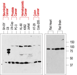
2. Anti-p75 NGF Receptor (extracellular) Antibody, preincubated with p75 NGF Receptor (extracellular) Blocking Peptide (#BLP-NT007). Western blot analysis of normal rat tissue (right) and in human cancer cell lines (left):p75NTR is visualized with Anti-p75 NGF Receptor (extracellular) Antibody (#ANT-007), (1:200). Note that human cancer cell lines from hemapoietic origin show high p75NTR expression, while cell lines from prostate and colon cancer origin show lower levels. Interestingly, p75NTR from rat (right blot and the C6 cell line) and human (left blot) samples run with a different apparent MW, probably due to species-specific differential glycosylation.
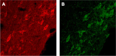
 Expression of p75NTR in rat brainImmunohistochemical staining of rat brain with Anti-p75 NGF Receptor (extracellular) Antibody (#ANT-007). A. Cells in the diagonal band are stained positive for p75NTR. B. Staining of the same section with goat anti-ChAT confirms that p75NTR staining is specific to cholinergic neurons.
 Expression of p75NTR in rat brainImmunohistochemical staining of rat brain with Anti-p75 NGF Receptor (extracellular) Antibody (#ANT-007). A. Cells in the nucleus basalis mangocellularis are stained positive for p75NTR. B. Staining of the same section with goat anti-ChAT confirms that the p75NTR staining is specific to cholinergic neurons.
Properties
Sequence
Accession (Uniprot) Number P08138
Peptide Confirmation Confirmed by amino acid analysis and mass spectrometry.
Purity >70%
Storage Before Reconstitution Lyophilized powder can be stored intact at room temperature for two weeks. For longer periods, it should be stored at -20°C.
Reconstitution 100 µl double distilled water (DDW).
Concentration After Reconstitution 0.4 mg/ml.
Storage After Reconstitution -20°C.
Antigen Preadsorption Control 1 µg peptide per 1 µg antibody.
Standard Quality Control Of Each Lot Western blot analysis.

