Overview
Type: Synthetic peptide
Form: Lyophilized powder
TRPV2/VRL1 (extracellular) Blocking Peptide (#BLP-CC039) is the original antigen used for immunization during Anti-TRPV2 (VRL1) (extracellular) Antibody (#ACC-039) generation. The blocking peptide binds and ‘blocks’ Anti-TRPV2/VRL1 (extracellular) primary antibody, this makes it a good negative reagent control to help confirm antibody specificity in western blot and immunohistochemistry applications. This control is also often called a pre-adsorption control.
Applications: wb, ihc
For research purposes only. not for human use
Applications
Demonstration of Pre-adsorption control
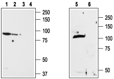 Western blot analysis of ND7/23 cell line membrane (lanes 1 and 3), RBL lysates (lanes 2 and 4) and rat brain membrane (lanes 5and 6):1,2,5. Anti-TRPV2 (VRL1) (extracellular) Antibody (#ACC-039), (1:200).
3,4,6. Anti-TRPV2 (VRL1) (extracellular) Antibody, preincubated with TRPV2/VRL1 (extracellular) Blocking Peptide (#BLP-CC039). Expression of TRPV2 in mouse DRGImmunohistochemical staining of TRPV2 in mouse dorsal root ganglion (DRG) using Anti-TRPV2 (VRL1) (extracellular) Antibody (#ACC-039). A. TRPV2 (green) appears in patches along the perimeter of the DRG (arrows). B. Neurons containing neurofilament 200 (red) are scattered in the DRG, also in patches (arrows). C. A merge of the two panels shows that the spatial distribution of neurofilament 200 and TRPV2 expression overlaps. However, DRGs showing robust expression of neurofilament 200 do not contain TRPV2.
 Western blot analysis of rat brain (lanes 1 and 3) and rat RBL basophilic leukemia cell lysate (lanes 2 and 4):1,2. Guinea pig Anti-TRPV2 (VRL1) (extracellular) Antibody (#ACC-039-GP), (1:2000).
3,4. Guinea pig Anti-TRPV2 (VRL1) (extracellular) Antibody, preincubated with TRPV2/VRL1 (extracellular) Blocking Peptide (#BLP-CC039). Multiplex staining of TRPV2 and mGluR5 in rat DRGImmunohistochemistry of rat dorsal root ganglion using Guinea pig Anti-TRPV2 (VRL1) (extracellular) Antibody (#ACC-039-GP) (1:60) and Anti-mGluR5 (extracellular)-ATTO Fluor-594 Antibody (#AGC-007-AR), (red), (1:60). A. TRPV2 staining (green). B. mGluR5 staining (red). C. Merge of A and B demonstrates co-localization of TRPV2 and mGluR5 in DRG cells. Nuclei are stained using DAPI as the counterstain (blue).
Properties
Sequence
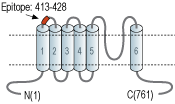
- (C)HQPSLDQPAIPSSKAT, corresponding to amino acid residues 413-428 of rat TRPV2 (Accession Q9WUD2).
Accession (Uniprot) Number Q9WUD2
Peptide Confirmation Confirmed by amino acid analysis and mass spectrometry.
Purity >70%
Storage Before Reconstitution Lyophilized powder can be stored intact at room temperature for two weeks. For longer periods, it should be stored at -20°C.
Reconstitution 100 µl double distilled water (DDW).
Concentration After Reconstitution 0.4 mg/ml.
Storage After Reconstitution -20°C.
Antigen Preadsorption Control 1 µg peptide per 1 µg antibody.
Standard Quality Control Of Each Lot Western blot analysis.
