A look at several excellent antibodies that target presynaptic and postsynaptic regions of the synapse.
While you can identify synapses with electron microscopy (EM), a lot of labs don’t have access to one of these and EM is usually a slow and relatively expensive procedure. A more accessible and rapid method is to label synapses with specific antibodies.
Here you can easily find multiple primary antibodies that will specifically target presynaptic or postsynaptic regions of the synapse. We make and test all our antibodies in-house, here at Alomone labs. If you’d like to know more about these antibodies, click on the link and head over the datasheet where you’ll find additional information, specific properties, and references.
Jump to
Immunohistochemistry protocol tips
There are a few things to keep in mind when you run immunohistochemistry (IHC) with synaptic markers. If you’re performing multiplex or colocalization studies, we highly recommend you consider using a primary antibody directly conjugated to a fluorophore. These eliminate the need for a secondary antibody and can overcome issues with finding compatible secondary antibodies against multiple species.
In some cases, antibody penetration can be lower than in other tissues. If you have this problem, you could try the following:
- Boosting Triton to 0.5% can improve penetration without noticeably compromising tissue integrity
- Increasing the tissue incubation to overnight (or even over several days) at 4oC may help
- Remove lipids with a clearing protocol like CLARITY or DISCO
For a complete step-by-step guide, take a look at our protocols pages that cover IHC, multiplex IHC, immunocytochemistry (ICC), and western blot (WB).
Presynaptic Markers
Antibodies for the presynaptic region include markers against major vesicle proteins found within the synapse, like synaptophysin, and amino acid transporters like the vesicular GABA transporter.
You can select individual markers, or you can get the Presynaptic Marker Antibody Kit (#AK-235), which contains all presynaptic markers at a lower price.
Neurexin-3α (membrane protein)
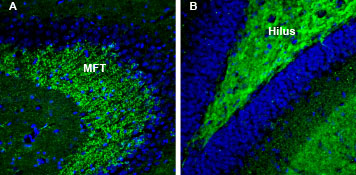
Neurexins (NRXNs) are a family of presynaptic transmembrane synaptic adhesion proteins expressed in all neurons. In the mammalian brain, they are known to interact with neuroligins, dystroglycan, and neurexophilins. Neurexin 3α splicing can yield secreted and transmembrane form.
Marker

SNAP-25 (cytoplasmic protein)
SNAP-25 (synaptosomal-associated 25kD protein) is a member of the soluble N-ethylmaleimide-sensitive factor attachment protein receptor (SNARE) protein superfamily. SNARE proteins play a crucial role in exocytosis and intracellular vesicle trafficking, making them essential for cell growth, hormone secretion, and neurotransmission.
Marker

Synapsin (cytoplasmic protein)
Synapsins are a family of synaptic vesicle-associated phosphoproteins that are important for fine-tuning synaptic transmission and remodeling. Synapsin I is a peripheral membrane protein of synaptic vesicles with a role in neurotransmitter release and cytoskeletal organization. Synapsin II, the collective name synapsin IIa and IIb, hep regulate synaptic function in the central and peripheral nervous system.
Markers
- Anti-Synapsin I (SYN1) Antibody (#ANR-014)
- Anti-Synapsin II (SYN2) Antibody (#ANR-015)
- Anti-Synapsin III (SYN3) Antibody (#ANR-017)


Synaptophysin (membrane protein)
Synaptophysin is part of a family of proteins, which includes synaptogyrin and synaptoporin, and is a major membrane protein found in the membrane of small synaptic vesicles.
Marker


Synaptotagmin-1 (cytoplasmic protein)
Synaptotagmins are type I membrane proteins usually found in intracellular vesicles and play a role in exocytosis and trafficking through Ca2+-dependent binding.
Marker

Syntaxin-1 (cytoplasmic protein)
Syntaxins belong to the SNARE protein superfamily and contribute to exocytosis. Syntaxin 1 is a type IV membrane protein with a relatively large N-terminus containing the SNARE motif found at the cytoplasmic side and a transmembrane domain.
Marker

VAMP-2 (cytoplasmic protein)
VAMP-2 (also known as synaptobrevin-2) is a member of the SNARE protein superfamily. VAMP-2 has been the subject of a lot of work thanks to its involvement with neuronal and neuroendocrine cell exocytosis where it functions as the vesicle membrane protein vesicular (v)-SNARE.
Marker

Vesicular GABA Transporter
The vesicular GABA transporter (VGAT) loads GABA and glycine into synaptic vesicles from the neuronal cytoplasm. VGAT expression is seen in synaptic vesicles of GABAergic and glycinergic neurons.
Marker
- Anti-Vesicular GABA Transporter (VGAT) Antibody (#AGT-005)
- Anti-Vesicular GABA Transporter (VGAT)-ATTO Fluor-594 Antibody (#AGT-005-AR)
- Guinea pig Anti-Vesicular GABA Transporter (VGAT) Antibody (#AGP-129)


Vesicular Glutamate Transporters (cytoplasmic proteins)
Vesicular Glutamate Transporters (VGLUTs) package and transport glutamate into synaptic vesicles. Three VGLUTs have been cloned: VGLUT1 (a brain-specific sodium-dependent inorganic phosphate (Pi) transporter) and its homologs VGLUT2 (differentiation-associated sodium-dependent Pi transporter) and VGLUT3. VGLUT1 and VGLUT2 expression is in cortical and subcortical neurons respectively. Non-glutamatergic neurons express VGLU3. VGLUT2 expression is in the thalamus, brainstem, and deep cerebellar nuclei.
Marker
- Anti-VGLUT1 Antibody (#AGC-035)
- Anti-VGLUT2 Antibody (#AGC-036)
- Anti-VGLUT2-ATTO Fluor-594 Antibody (#AGC-036-AR)
- Anti-VGLUT3 Antibody (#AGC-037)


Growth-Associated Protein-43
Growth-associated protein 43 (GAP43) is a neuron-specific phosphoprotein that plays an important role in nerve growth, development, regeneration, and axonal remodeling. You can find GAP43 expression in developing neurons, especially in the growth cone. In mature neurons, GAP43 is seen in regenerating nerves.
Marker

Vesicular Monoamine Transporters
Vesicular monoamine transporters (VMATs) are found in the membrane of presynaptic vesicles and are essential for proper monoaminergic neurotransmission, which requires the sequestration of the transmitter into synaptic vesicles by VMAT for subsequent Ca2+-stimulated exocytotic release.
Markers


Postsynaptic Markers
Postsynaptic markers include scaffolding proteins like gephyrin and the shank family, as well as cell-surface proteins like neuroligins.
Just like with the presynaptic markers, can select individual postsynaptic markers, or you can purchase the Postsynaptic Marker Antibody Kit (#AK-236), which contains all postsynaptic markers at a lower price.
Neuroligin (membrane protein)
Neuroligins act as ligands for β-neurexins. Neuroligin and β-neurexin “shake hands,” which forms the connection between two neurons. Neuroligin 1 may help form and remodel synapses in the central nervous system (CNS), while neuroligin 2 helps promote synaptic maturation.
Markers
- Anti-Neuroligin 1 (extracellular) Antibody (#ANR-035)
- Anti-Neuroligin 2 (extracellular) Antibody (#ANR-036)


Postsynaptic Density-95
The postsynaptic density (PSD) comprises membrane-associated proteins that specialize in postsynaptic signal transduction and processing. PSD-95 interacts with both ionotropic and metabotropic glutamate receptors via protein-protein interactions and plays a role in their assembly and spatial organization as well as coupling these receptors to downstream signaling events.
Marker

Synapse-Associated Protein-102
Synapse-associated protein (SAP)-102 is a part of the PSD, a dedicated structure formed in postsynaptic nerve terminals that includes a specialized assembly of ion channels, receptors, and signaling molecules involved in information processing and the modulation of synaptic plasticity. SAP-102 also interacts with NMDA receptor complexes.
Marker

Shank
Shank family proteins (shank1, shank2, and shank3) are neuronal scaffold proteins that organize a cytoskeleton-associated signaling complex at the PSD of nearly all excitatory glutamatergic synapses in the mammalian brain.
Markers


Homer1
The Homer protein family includes Homer1, 2, and 3. These proteins are mainly in the neuronal PSD where they play a role in post-synaptic signaling and receptor trafficking by forming multivalent links with various receptors and PSD scaffolding proteins. The homer protein family has three members with each member alternatively spliced into long and short forms.
Marker


Gephyrin (cytoplasmic protein)
Gephyrin is a highly conserved, intracellular scaffolding protein responsible for the post-synaptic anchoring and assembly of the inhibitory glycine and γ-aminobutyric acid type A receptors (GlyR and GABAAR).
Marker

Still need helping to choose the best synaptic markers? Speak with our scientific advisors and we’ll make sure you find what’s best for your research.
